Mycobacterium tuberculosis-induced expression of granulocyte-macrophage colony stimulating factor is mediated by PI3-K/MEK1/p38 MAPK signaling pathway
- PMID: 23615263
- PMCID: PMC4133881
- DOI: 10.5483/bmbrep.2013.46.4.200
Mycobacterium tuberculosis-induced expression of granulocyte-macrophage colony stimulating factor is mediated by PI3-K/MEK1/p38 MAPK signaling pathway
Abstract
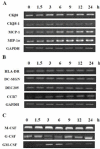
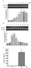
Members of the colony stimulating factor cytokine family play important roles in macrophage activation and recruitment to inflammatory lesions. Among them, granulocyte-macrophage colony stimulating factor (GM-CSF) is known to be associated with immune response to mycobacterial infection. However, the mechanism through which Mycobacterium tuberculosis (MTB) affects the expression of GM-CSF is poorly understood. Using PMA-differentiated THP-1 cells, we found that MTB infection increased GM-CSF mRNA expression in a dosedependent manner. Induction of GM-CSF mRNA expression peaked 6 h after infection, declining gradually thereafter and returning to its basal levels at 72 h. Secretion of GM-CSF protein was also elevated by MTB infection. The increase in mRNA expression and protein secretion of GM-CSF caused by MTB was inhibited in cells treated with inhibitors of p38 MAPK, mitogen-activated protein kinase kinase (MEK-1), and PI3-K. These results suggest that up-regulation of GM-CSF by MTB is mediated via the PI3-K/MEK1/p38 MAPK-associated signaling pathway.
Figures


Similar articles
-
Cytokine-specific activation of distinct mitogen-activated protein kinase subtype cascades in human neutrophils stimulated by granulocyte colony-stimulating factor, granulocyte-macrophage colony-stimulating factor, and tumor necrosis factor-alpha.Blood. 1999 Jan 1;93(1):341-9. Blood. 1999. PMID: 9864179
-
Effects of GM-CSF and M-CSF on tumor progression of lung cancer: roles of MEK1/ERK and AKT/PKB pathways.Int J Mol Med. 2006 Aug;18(2):365-73. Int J Mol Med. 2006. PMID: 16820947
-
Mechanisms of Chlamydophila pneumoniae-mediated GM-CSF release in human bronchial epithelial cells.Am J Respir Cell Mol Biol. 2006 Mar;34(3):375-82. doi: 10.1165/rcmb.2004-0157OC. Epub 2005 Dec 9. Am J Respir Cell Mol Biol. 2006. PMID: 16340003
-
c-Jun N-terminal kinase (JNK) and p38 mitogen-activated protein kinase (p38 MAPK) are involved in Mycobacterium tuberculosis-induced expression of Leukotactin-1.BMB Rep. 2012 Oct;45(10):583-8. doi: 10.5483/bmbrep.2012.45.10.120. BMB Rep. 2012. PMID: 23101513
-
Activation of mitogen-activated protein kinase cascades during priming of human neutrophils by TNF-alpha and GM-CSF.J Leukoc Biol. 1998 Oct;64(4):537-45. J Leukoc Biol. 1998. PMID: 9766635
Cited by
-
Functional roles of Syk in macrophage-mediated inflammatory responses.Mediators Inflamm. 2014;2014:270302. doi: 10.1155/2014/270302. Epub 2014 Jun 18. Mediators Inflamm. 2014. PMID: 25045209 Free PMC article. Review.
-
Identifcation of differentially expressed long non-coding RNAs in CD4+ T cells response to latent tuberculosis infection.J Infect. 2014 Dec;69(6):558-68. doi: 10.1016/j.jinf.2014.06.016. Epub 2014 Jun 27. J Infect. 2014. PMID: 24975173 Free PMC article.
-
Evaluation of cytokines in peripheral blood mononuclear cell supernatants for the diagnosis of tuberculosis.J Inflamm Res. 2018 Dec 24;12:15-22. doi: 10.2147/JIR.S183821. eCollection 2019. J Inflamm Res. 2018. PMID: 30636888 Free PMC article.
-
Anti-oxidizing effect of the dichloromethane and hexane fractions from Orostachys japonicus in LPS-stimulated RAW 264.7 cells via upregulation of Nrf2 expression and activation of MAPK signaling pathway.BMB Rep. 2014 Feb;47(2):98-103. doi: 10.5483/bmbrep.2014.47.2.088. BMB Rep. 2014. PMID: 24219867 Free PMC article.
-
GM-CSF targeted immunomodulation affects host response to M. tuberculosis infection.Sci Rep. 2018 Jun 5;8(1):8652. doi: 10.1038/s41598-018-26984-3. Sci Rep. 2018. PMID: 29872095 Free PMC article.
References
-
- World Health Organization. WHO report 2007: Global tuberculosis control; surveillance, planning, financing. WHO; Geneva: 2007. pp. 1–277.
-
- World Health Organization. WHO report 2007: Global MDR-TB and XDR-TB response plan 2007-2008. WHO; Geneva: 2007. pp. 1–48.
Publication types
MeSH terms
Substances
LinkOut - more resources
Full Text Sources
Other Literature Sources
Miscellaneous

