. 2013 Jun;24(12):1933-46.
doi: 10.1091/mbc.E12-12-0851. Epub 2013 Apr 24.

Activation of ADF/cofilin by phosphorylation-regulated Slingshot phosphatase is required for the meiotic spindle assembly in Xenopus laevis oocytes

Affiliations

Activation of ADF/cofilin by phosphorylation-regulated Slingshot phosphatase is required for the meiotic spindle assembly in Xenopus laevis oocytes

Shohei Iwase et al. Mol Biol Cell. 2013 Jun.

Abstract

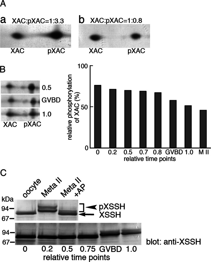
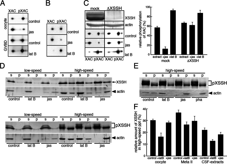
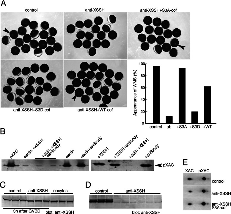
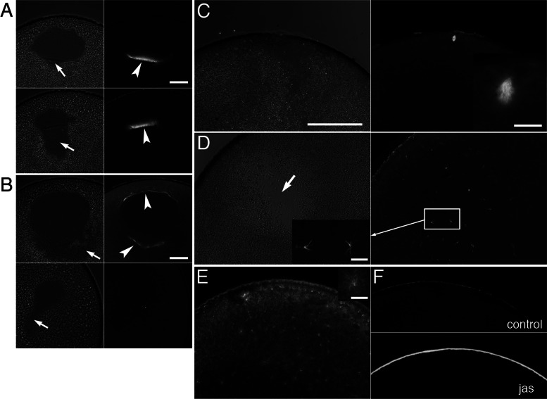
We identify Xenopus ADF/cofilin (XAC) and its activator, Slingshot phosphatase (XSSH), as key regulators of actin dynamics essential for spindle microtubule assembly during Xenopus oocyte maturation. Phosphorylation of XSSH at multiple sites within the tail domain occurs just after germinal vesicle breakdown (GVBD) and is accompanied by dephosphorylation of XAC, which was mostly phosphorylated in immature oocytes. This XAC dephosphorylation after GVBD is completely suppressed by latrunculin B, an actin monomer-sequestering drug. On the other hand, jasplakinolide, an F-actin-stabilizing drug, induces dephosphorylation of XAC. Effects of latrunculin B and jasplakinolide are reconstituted in cytostatic factor-arrested extracts (CSF extracts), and XAC dephosphorylation is abolished by depletion of XSSH from CSF extracts, suggesting that XSSH functions as an actin filament sensor to facilitate actin filament dynamics via XAC activation. Injection of anti-XSSH antibody, which blocks full phosphorylation of XSSH after GVBD, inhibits both meiotic spindle formation and XAC dephosphorylation. Coinjection of constitutively active XAC with the antibody suppresses this phenotype. Treatment of oocytes with jasplakinolide also impairs spindle formation. These results strongly suggest that elevation of actin dynamics by XAC activation through XSSH phosphorylation is required for meiotic spindle assembly in Xenopus laevis.


Figures

FIGURE 1:
FIGURE 1:
Phosphorylation states of XAC and XSSH during oocyte maturation. (A) Phosphorylation states of XAC as determined by 2D immunoblots in immature oocytes (a) and mature (Meta II) oocytes (b). Unphosphorylated and phosphorylated forms of XAC are indicated by XAC and pXAC, respectively. Relative amounts of XAC and pXAC were determined by densitometry and are indicated on top of each blot. (B) Time course of changes in phosphorylation levels of XAC during oocyte maturation. Left, three examples of 2D immunoblots of XAC at relative time points as indicated in the graph on the right. The spots of XAC and pXAC at different time points during oocyte maturation were quantified by densitometry, and percentages of pXAC are shown. Time 0 is at the addition of progesterone, and time 1.0 is when >80% of oocytes show WMS. GVBD represents the oocytes in which GVBD just occurred. M II represents the data from fully mature oocytes. (C) Phosphorylation states of XSSH during oocyte maturation as determined by mobility shifts in immunoblot. Top, immunoblots of XSSH in immature (oocyte) and mature (Meta II) oocytes. XSSH in mature oocyte were detected as bands with slower mobility than those of XSSH in immature oocytes. The retarded mobility was canceled by alkaline phosphatase treatment (Meta II + AP), indicating that the mobility shifts were due to phosphorylation. The bands of pXSSH were split into two groups. Bottom, immunoblots of XSSH at different time points during oocyte maturation. Phosphorylation of XSSH as shown by mobility shifts was enhanced at GVBD.
FIGURE 2:
FIGURE 2:
Identification of phosphorylation sites of XSSH and binding properties of XSSH to 14-3-3ζ. (A) Domain organization of XSSH and structures of truncated mutants used in this study. SSH a and SSH b, Slingshot-homology domains A and B, respectively; DSPc, a dual-specific phosphatase domain; Tail, Slingshot tail domain. (B, C) Phosphorylation of XSSH and the mutants with CSF extracts. (a–a′′′) treatments with buffer alone, (b–b′′′) treatments with CSF extracts, and (c–c′′′) treatments with CSF extracts followed by alkaline phosphatase treatments. Arrowheads represent shifted bands due to phosphorylation, and arrows indicate the dephosphorylated forms after alkaline phosphatase treatments. Because samples were probed with the anti-GST antibody, degradation products were also detected. Phosphorylation of the degraded GST–N-tail was also detected, as shown by a bracket in C. (D) In vitro phosphorylation of GST–N-tail was performed in buffer alone (control), meiotic extracts (CSF), interphase extracts (interphase), or mitotic extracts (ΔN cyclin) and subjected to immunoblot with anti-GST antibody. In the two right lanes, GST–N-tail was reacted with CSF extracts (CSF) or CSF extracts with 1 μM okadaic acid (CSF+OKA). Arrowhead indicates the position of phosphorylated bands, and an arrow indicates the position of additionally shifted bands in the presence of okadaic acid. (E) Phosphorylation sites in the N-tail region. The yellow boxes indicate phosphorylated residues identified by MS. P1–P5 represent residues examined by site-directed mutagenesis. The underlining at the P4 site indicates the consensus sequence for PKD1 phosphorylation sites. (F) GST–N-tail (pGST–N-tail) and its variants with indicated mutations (P1,3 to P1,2,3,4,5) were phosphorylated in vitro in CSF extracts and subjected to immunoblot with anti-GST antibody. Unphosphorylated GST–N-tail was loaded on the right lane. Arrow and arrowhead indicate nonphosphorylated and fully phosphorylated GST–N-tail, respectively. (G) Pull-down assay of 14-3-3ζ with GST-tail mutants. 14-3-3ζ bound to GST-tail (left) and GST-C-tail (right) before and after phosphorylation but did not bind to GST-N-tail (right). Top right, immunoblots with anti-GST antibody of nonphosphorylated and phosphorylated GST-tail mutants bound to glutathione beads. GST alone was used as control. Arrowheads indicate phosphorylated GST–N-tail and GST-C-tail. Bottom right, coprecipitated 14-3-3ζ probed with anti–14-3-3 antibody.
FIGURE 3:
FIGURE 3:
Effects of phosphorylation of XSSH on its phosphatase activity (A, D) and F-actin–binding ability (B, C) in vitro. (A) Time course of dephosphorylation of 10 μM pXAC by 0.2 μM XSSH in the absence of F-actin (control), 0.2 μM XSSH in the presence of 4.2 μM F-actin (XSSH), or 0.2 μM pXSSH in the presence of 4.2 μM F-actin (pXSSH). Dephosphorylation of pXAC was monitored by immunoblots using anti–phospho-ADF/cofilin antibody (top left, three). The results were quantified by densitometry and are shown as relative band intensity in the graph (1.0 at time 0). Bottom left two, time course of dephosphorylation of pXAC under the same conditions as described except for the presence of 4.7 μM chicken cofilin with 4.2 μM F-actin (XSSH+cof and pXSSH+cof). (B) Pelleting assay of XSSH (top) and pXSSH (bottom) with 4.7 μM F-actin by altering the concentrations of XSSH or pXSSH as indicated. Supernatants (s) and pellets (p) were examined by SDS–PAGE and Coomassie staining. The graph shows concentration of XSSHs in pellets vs. that of added XSSHs. The data from three independent experiments are plotted. (C) Pelleting assay of 1 μM XSSH or pXSSH with 4.7 μM F-actin in the presence (+cofilin) or absence (–) of 4.7 μM chick cofilin.
FIGURE 4:
FIGURE 4:
Effects of F-actin levels on the XAC phosphatase activity of XSSH. (A) Phosphorylation states of XAC were examined by 2D immunoblots of XAC in immature oocytes (oocyte) and maturing oocytes just after GVBD, which were treated with jasplakinolide (jas) or latrunculin B (lat B). Control indicates the treatment with vehicle alone. (B) Phosphorylation states of XAC were examined by 2D immunoblots of XAC in CSF extracts in the presence of jas or lat B. Control indicates addition of vehicle alone. (C) Phosphorylation states of XAC were examined by 2D immunoblots of XAC in mock-depleted CSF extracts (mock) or XSSH-depleted CSF extracts (ΔXSSH) in the absence (control) or presence of jas or lat B. XSSH was sufficiently depleted from CSF extracts (front row, XSSH) as judged by the anti-actin blots (second row, actin). The spots of XAC and pXAC were quantified by densitometry, and percentages of pXAC are shown. (D) Immature oocytes (top) and maturing oocytes just after GVBD (bottom) treated with vehicle alone (control), lat B, or jas were centrifuged at 12,000 × g for 15 min (low speed), and the supernatants were further ultracentrifuged at 436,000 × g for 20 min (high speed) to separate the supernatants and pellets. The supernatants (s) and pellets (p) were subjected to SDS–PAGE and examined by immunoblotting with anti-XSSH or anti-actin antibody. (E) Immunoblots of XSSH and actin in high-speed supernatants (s) and pellets (p) of CSF extracts treated with vehicle alone (control), 25 μM lat B, 10 μM jas, or 10 μM phalloidin (pha) as described in D. (F) The XSSH bands in high-speed supernatants and precipitates (ppt) were quantified by densitometry, and percentages of precipitated XSSH are shown. Bars, mean + SE from three independent experiments using immature oocytes (oocyte), mature oocytes (Meta II), and CSF extracts in the absence (control) or presence of latrunculin B (+latB) or jasplakinolide (+jas).
FIGURE 5:
FIGURE 5:
Effects of the anti-XSSH antibody on the XSSH activity in vitro and the phosphorylation states of XSSH and XAC in maturing oocytes. (A) Xenopus oocytes were injected with buffer alone (control), anti-XSSH, anti-XSSH plus constitutively active cofilin (S3A-cof), anti-XSSH plus phosphomimetic inactive cofilin (S3D-cof), or anti-XSSH plus wild-type cofilin (WT-cof). The concentrations of anti-XSSH IgG and cofilins were 8 and 0.5 mg/ml, respectively. After progesterone treatment, WMS formation (indicated by arrowheads) was examined. A typical example of injection experiments is summarized in the graph. ab, anti-XSSH antibody injection. (B) Effect of the anti-XSSH antibody on the XAC phosphatase activity of XSSH was examined by incubating pXAC (left lane) with combinations of 4.7 μM F-actin, 0.3 μM XSSH, and 1 mg/ml antibody as indicated, followed by immunoblot with anti–phospho-ADF/cofilin antibody. pXAC was dephosphorylated in the presence of F-actin and XSSH, and addition of anti-XSSH antibody did not inhibit dephosphorylation. Actin alone, antibody alone, XSSH alone, or actin and the antibody without XSSH did not alter pXAC dephosphorylation. The anti-XSSH antibody did not activate XSSH in the absence of F-actin. (C) Effects of anti-XSSH antibody on phosphorylation of XSSH in oocytes. Oocytes were injected with buffer alone (control; three samples) or anti-XSSH antibody (three samples) and examined at 3 h after GVBD by immunoblot with anti-XSSH antibody. Right, three samples of untreated immature oocytes as controls to determine mobility of nonphosphorylated XSSH. Injection of anti-XSSH antibody partially inhibited full phosphorylation of XSSH during oocyte maturation. (D) Binding of the anti-XSSH antibody to XSSH in mature oocytes. Control and antibody-injected oocytes were homogenized with PBS and centrifuged (at low speed). The supernatants were subjected to SDS–PAGE and immunoblotting with anti-XSSH antibody. Three control and six antibody-injected samples are shown. (E) Effects of the anti-XSSH antibody on the phosphorylation states of XAC during oocyte maturation. Oocytes were injected with buffer alone (control), anti-XSSH antibody, or anti-XSSH antibody and S3A-cofilin and examined by 2D immunoblot with anti-XAC antibody, when >80% of control oocytes show WMS.
FIGURE 6:
FIGURE 6:
Effects of injection of anti-XSSH antibody on assembly of MTOC-TMA and meiotic spindles. (A, B) Phase contrast (left) and immunofluorescence (right) microscopy of control oocytes injected with buffer alone (A) or oocytes injected with anti-XSSH antibody (B). Microtubules were stained with anti-tubulin antibody (right). Assembly of MTOC-TMA (arrowhead in A, top right) at the basal region of GV and its migration toward the animal pole (arrowhead in A, bottom right) are clearly visible in control (A), whereas its structure is faint in antibody-injected oocytes (arrowheads in B, top right). The yolk-free region formed at the basal region of GV (indicated by arrows in phase contrast micrographs) showed broader and irregular shape in anti-XSSH antibody-injected oocytes (B). (C, D) Phase contrast images (left) and immunofluorescence staining for tubulin (right) of control oocytes 4 h after GVBD (C) and antibody-injected oocytes fixed at 4 h after control oocytes formed WMS (D). Meiotic spindles were already assembled in control oocytes (C, magnified in inset), whereas fine and radial microtubule structures were formed (D, left, magnified) and scattered around the yolk-free region in antibody-injected oocytes (D, arrow). (E) A jasplakinolide-treated oocyte was fixed at 4 h after control oocytes formed WMS and stained with anti-tubulin antibody. An example of radial microtubule structures is shown in the inset on the upper right corner. (F) Staining of control (top) and jasplakinolide-treated (bottom) oocytes for actin filaments by anti-actin antibody. Note the cortical accumulation of actin filaments by jasplakinolide treatment. Bars, (A, B) 100 μm, (C, applies to C– F) 200 μm, (insets) 20 μm.

Similar articles

Cited by

References

    1. Abe H, Obinata T, Minamide LS, Bamburg JR. Xenopus laevis actin-depolymerizing factor/cofilin: a phosphorylation-regulated protein essential for development. J Cell Biol. 1996;132:871–885. - PMC - PubMed
    1. Arber S, Barbayannis FA, Hanser H, Schneider C, Stanyon CA, Bernard O, Caroni P. Regulation of actin dynamics through phosphorylation of cofilin by LIM-kinase. Nature. 1998;393:805–809. - PubMed
    1. Azoury J, Lee KW, Georget V, Hikal P, Verlhac MH. Symmetry breaking in mouse oocytes requires transient F-actin meshwork destabilization. Development. 2011;138:2903–2908. - PubMed
    1. Azoury J, Lee KW, Georget V, Rassinier P, Leader B, Verlhac MH. Spindle positioning in mouse oocytes relies on a dynamic meshwork of actin filaments. Curr Biol. 2008;18:1514–1519. - PubMed
    1. Barišicˇ S, Nagel AC, Franz-Wachtel M, Macek B, Preiss A, Link G, Maier D, Hausser A. Phosphorylation of Ser 402 impedes phosphatase activity of slingshot 1. EMBO Rep. 2011;12:527–533. - PMC - PubMed

Publication types

MeSH terms

LinkOut - more resources