. 2013 Apr 24;14(5):8787-800.
doi: 10.3390/ijms14058787.

The cloning and characterization of the enolase2 gene of Gekko japonicus and its polyclonal antibody preparation

Affiliations

The cloning and characterization of the enolase2 gene of Gekko japonicus and its polyclonal antibody preparation

Jing Li et al. Int J Mol Sci. .

Abstract

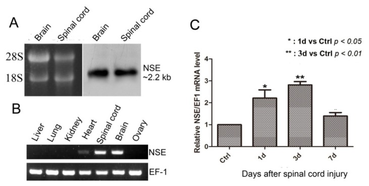
The enolase2 gene is usually expressed in mature neurons and also named neuron specific enolase (NSE). In the present study, we first obtained the NSE gene cDNA sequence by using the RACE method based on the expressed sequence tag (EST) fragment from the cDNA library of Gekko japonicus and identified one transcript of about 2.2 kb in central nervous system of Gekko japonicus by Northern blotting. The open reading frame of NSE is 1305 bp, which encodes a 435 amino-acid protein. We further investigated the multi-tissue expression pattern of NSE by RT-PCR and found that the expression of NSE mRNA was very high in brain, spinal cord and low in heart, while it was not detectable in other tissues. The real-time quantitative PCR was used to investigate the time-dependent change in the expression of the NSE mRNA level after gecko spinal cord transection and found it significantly increased at one day, reaching its highest level three days post-injury and then decreasing at the seventh day of the experiment. The recombinant plasmid of pET-32a-NSE was constructed and induced to express His fused NSE protein. The purified NSE protein was used to immunize rabbits to generate polyclonal antisera. The titer of the antiserum was more than 1:65536 determined by ELISA. Western blotting showed that the prepared antibody could specifically recognize the recombinant and endogenous NSE protein. The result of immunohistochemistry revealed that positive signals were present in neurons of the brain and the spinal cord. This study provided the tools of cDNA and polyclonal antibody for studying NSE function in Gekko japonicus.


Figures

Figure 1
Figure 1
Molecular cloning and bioinformatic analysis of NSE. (A) The NSE cDNA (GenBank accession No. JQ080314) and its deduced amino acid sequence. The full-length cDNA of NSE was 1305 bp and the open reading frame encoded a polypeptide of 434 amino acids. The primers of RACE are boxed. The numbering of the nucleotide and amino acid sequences is shown on the right; (B) alignment of NSE using the MegAlign program (DNASTAR) by the CLUSTAL method. Shaded (with solid black) residues are the amino acids that match the consensus. NSE amino acid sequences were obtained from previously reported sequences in GenBank, including Danio rerio, Bos taurus, Gallus gallus, Mus musculus, Rattus norvegicus and Homo sapiens; (C) phylogenetic tree analysis of NSE gene from Gekko japonicus and other species was constructed by neighbor-joining methods within the package PHYLIP 3.5c. Bootstrap majority consensus values on 1000 replicates are indicated at each branch.
Figure 2
Figure 2
NSE mRNA transcript in CNS, the expression pattern in different tissues and mRNA expression after spinal cord transection in geckos. (A) The representative result of Northern blotting analyses of NSE mRNA in CNS of adult gecko. Ten micrograms of total RNA samples from brain and spinal cord of gecko were separated in 1% formaldehyde-denatured (w/v) agarose gel (Left panel), with the 18S and 28S rRNAs of corresponding tissues shown on the left. The 680 bp from NSE-open read frame (ORF) sequence was obtained by the PCR method and cloned into the pGEM-T Easy vector. Dig-labeled NSE riboprobes were synthesized in vitro from linearized plasmid above with T7 RNA polymerase. The hybridized bands were visualized with CDP-Star chemiluminescent substrate and recorded by X-ray film. The result of Northern blotting (Right panel) showed the length of NSE mRNA about 2.2 kb; (B) the representative results of semi-quantitative RT-PCR analyses of NSE mRNA in the different tissues of adult geckos, including liver, lung, kidney, heart, spinal cord, brain and ovary of adult geckos. The results revealed that the expression level of NSE mRNA was high in brain, spinal cord and low in heart, while it was not detectable in other tissues; (C) real-time qPCR analysis of NSE expression in the spinal cord after transection for the controls (Ctrl) and one, three and seven days post-injury. EF-1α was used for the quantitative normalization. * 1 d vs. Ctrl, p < 0.05; ** 3 d vs. Ctrl, p < 0.01.
Figure 3
Figure 3
Expression, purification of His fusion NSE protein and the titer of the prepared antiserum. (A) Expression of His tagged NSE protein with isopropyl β-d-1-thiogalactopyranoside (IPTG) induction. Lane 1 and 5: the total protein of uninduced pET-32a vector and pET-32a-gNSE plasmid. Lane 2–4: the induced total protein of pET-32a vector with 0.1 mM, 0.5 mM and 1.0 mM IPTG, respectively. The bands showed His tag protein of pET-32a vector was about 17.5 kDa (↓) and the same expression efficiency at different IPTG concentration. Lane 6–8: the induced total protein of pET-32a-gNSE plasmid with 0.1 mM, 0.5 mM, 1.0 mM IPTG, respectively. The bands showed His fusion protein of pET-32a-gNSE plasmid was about 67 kDa (←) and the same expression efficiency at different IPTG concentration. Lane M showed the standard MW protein and their MW list on the left of the Lane 1, respectively; (B) Lane 1(↓) showed the result of purification of His fused gecko NSE protein. The Lanes of 2 to 4 were the BSA, protein and their loading amounts were 1 μg, 2 μg and 3 μg, respectively. Lane M showed the standard MW protein and their MW list on the left of the Lane 1, respectively. Staining assay using 10% SDS-PAGE and Coomassie brilliant blue R-250 was applied to assess the induced expression and the result of purification of the recombinant fusion protein; (C) Titer of the prepared antiserum was determined by ELISA. The blue curve showed the absorbance values of antiserum at different dilutions and the purple of pre-immune serum at the same dilution gradient.
Figure 3
Figure 3
Expression, purification of His fusion NSE protein and the titer of the prepared antiserum. (A) Expression of His tagged NSE protein with isopropyl β-d-1-thiogalactopyranoside (IPTG) induction. Lane 1 and 5: the total protein of uninduced pET-32a vector and pET-32a-gNSE plasmid. Lane 2–4: the induced total protein of pET-32a vector with 0.1 mM, 0.5 mM and 1.0 mM IPTG, respectively. The bands showed His tag protein of pET-32a vector was about 17.5 kDa (↓) and the same expression efficiency at different IPTG concentration. Lane 6–8: the induced total protein of pET-32a-gNSE plasmid with 0.1 mM, 0.5 mM, 1.0 mM IPTG, respectively. The bands showed His fusion protein of pET-32a-gNSE plasmid was about 67 kDa (←) and the same expression efficiency at different IPTG concentration. Lane M showed the standard MW protein and their MW list on the left of the Lane 1, respectively; (B) Lane 1(↓) showed the result of purification of His fused gecko NSE protein. The Lanes of 2 to 4 were the BSA, protein and their loading amounts were 1 μg, 2 μg and 3 μg, respectively. Lane M showed the standard MW protein and their MW list on the left of the Lane 1, respectively. Staining assay using 10% SDS-PAGE and Coomassie brilliant blue R-250 was applied to assess the induced expression and the result of purification of the recombinant fusion protein; (C) Titer of the prepared antiserum was determined by ELISA. The blue curve showed the absorbance values of antiserum at different dilutions and the purple of pre-immune serum at the same dilution gradient.
Figure 4
Figure 4
The representative results of Western blot and immunochemical analyses. (A) Lane 1 and 2 showed the result of Western blot by using the generated rabbit polyclonal antibody against His tagged gecko NSE protein and the commercial monoclonal antibody against His protein, both bands showed the MW of His-tagged gecko NSE protein was about 67 kDa. The right panel showed the result of Western blot by using the generated rabbit polyclonal antibody against NSE protein obtained from gecko liver, kidney, brain and spinal cord, and the bands were about 47 kDa. The beta-actin was used as the internal control; (B) The immunochemical assay showed specificity of the polyclonal antibody on the gecko spinal cord (a) and brain (b) sections. NSE was expressed in cytoplasm of mature neurons, and the fluorescence signals were mainly distributed in grey matter of spinal cord and extensively detected in brain.

Similar articles

Cited by

References

    1. Chernoff E.A. Spinal cord regeneration: A phenomenon unique to urodeles? Int. J. Dev. Biol. 1996;40:823–831. - PubMed
    1. Goldshmit Y., Sztal T.E., Jusuf P.R., Hall T.E., Nguyen-Chi M., Currie P.D. Fgf-dependent glial cell bridges facilitate spinal cord regeneration in zebrafish. J. Neurosci. 2012;32:7477–7492. - PMC - PubMed
    1. Romero-Aleman M.M., Monzon-Mayor M., Yanes C., Lang D. Radial glial cells, proliferating periventricular cells, and microglia might contribute to successful structural repair in the cerebral cortex of the lizard Gallotia galloti. Exp. Neurol. 2004;188:74–85. - PubMed
    1. Dallimore E.J., Park K.K., Pollett M.A., Taylor J.S., Harvey A.R. The life, death and regenerative ability of immature and mature rat retinal ganglion cells are influenced by their birthdate. Exp. Neurol. 2010;225:353–365. - PubMed
    1. Brockes J.P. Amphibian limb regeneration: Rebuilding a complex structure. Science. 1997;276:81–87. - PubMed

Publication types

MeSH terms

Associated data

LinkOut - more resources