. 2013 May 16;497(7449):369-73.
doi: 10.1038/nature12069. Epub 2013 Apr 24.

Protective astrogenesis from the SVZ niche after injury is controlled by Notch modulator Thbs4

Affiliations

Protective astrogenesis from the SVZ niche after injury is controlled by Notch modulator Thbs4

Eric J Benner et al. Nature. .

Abstract

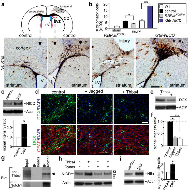
Postnatal/adult neural stem cells (NSCs) within the rodent subventricular zone (SVZ; also called subependymal zone) generate doublecortin (Dcx)(+) neuroblasts that migrate and integrate into olfactory bulb circuitry. Continuous production of neuroblasts is controlled by the SVZ microenvironmental niche. It is generally thought that enhancing the neurogenic activities of endogenous NSCs may provide needed therapeutic options for disease states and after brain injury. However, SVZ NSCs can also differentiate into astrocytes. It remains unclear whether there are conditions that favour astrogenesis over neurogenesis in the SVZ niche, and whether astrocytes produced there have different properties compared with astrocytes produced elsewhere in the brain. Here we show in mice that SVZ-generated astrocytes express high levels of thrombospondin 4 (Thbs4), a secreted homopentameric glycoprotein, in contrast to cortical astrocytes, which express low levels of Thbs4. We found that localized photothrombotic/ischaemic cortical injury initiates a marked increase in Thbs4(hi) astrocyte production from the postnatal SVZ niche. Tamoxifen-inducible nestin-creER(tm)4 lineage tracing demonstrated that it is these SVZ-generated Thbs4(hi) astrocytes, and not Dcx(+) neuroblasts, that home-in on the injured cortex. This robust post-injury astrogenic response required SVZ Notch activation modulated by Thbs4 via direct Notch1 receptor binding and endocytosis to activate downstream signals, including increased Nfia transcription factor expression important for glia production. Consequently, Thbs4 homozygous knockout mice (Thbs4(KO/KO)) showed severe defects in cortical-injury-induced SVZ astrogenesis, instead producing cells expressing Dcx migrating from SVZ to the injury sites. These alterations in cellular responses resulted in abnormal glial scar formation after injury, and significantly increased microvascular haemorrhage into the brain parenchyma of Thbs4(KO/KO) mice. Taken together, these findings have important implications for post-injury applications of endogenous and transplanted NSCs in the therapeutic setting, as well as disease states where Thbs family members have important roles.


Figures

Figure 1
Figure 1. SVZ generation of Thbs4hi astrocytes
a, Western blot analysis of Thbs4 protein levels in differentiated primary SVZ and cortical (ctx) astrocyte cultures. b, qPCR analyses of Thbs4 levels in FACS-sorted SVZ vs. ctx GFP+ astrocytes from GFAP-GFP transgenic mice, * p < 0.001, n = 5, Student's t-test; error bars = sem. c, Schematic representation of cortical transplantation strategy. In lower panels, Thbs4, tdTomato, GFAP IHC antibody staining of brain sections, 2-4 weeks after animals were transplanted with lineage-traced primary SVZ NSC cultures derived from tamoxifen-induced nestin-CreERtm4; r26r-tdTomato (N4; RTM) animals, showing co-localization between tdTomato, Thbs4, GFAP (arrowheads). Scale bar: 20 μm (c).
Figure 2
Figure 2. Thbs4hi astrocyte production following photothrombotic cortical injury
a, Schematic representation of photothrombosis injury model, with dashed box indicating region of imaging in (b). b, DAB IHC staining for tdTomato from N4; RTM animals induced with tamoxifen, showing a delayed activation of lineage-traced tdTomato+ cells to injury site 14 dpi. LV = lateral ventricle. c, Coronal sections of injury site 14 dpi, IHC stained for GFAP, Thbs4, and tdTomato, showing that lineage-traced tdTomato+ cells adjacent to injury are Thbs4hiGFAP+ astrocytes (arrowheads). d, Quantitative analyses of total tdTomato+ cells at injury site expressing Thbs4 14 dpi (88.10 ± 1.99% stdev, n = 3 animals). e, Western blot and quantitative analyses of Thbs4 protein levels in SVZ tissues 3dpi. * p < 0.001, n = 5, Student's t-test, error bar = sem. f, DAB IHC staining for Thbs4 expression 3 dpi, Nissl-counterstained. Ipsilateral and contralateral SVZ from the same brain section, imaged under identical conditions. Scale bars: 100 μm (b), 20 μm (c), 50 μm (f).
Figure 3
Figure 3. Notch signaling and regulation of injury-induced SVZ astrogenesis
a, Photothrombosis cortical injury model, with areas of imaging indicated by dashed boxes. Representative sections 14 dpi, DAB IHC staining for tdTomato from tamoxifen-induced N4; RTM; RBPJkFlox/+ (control); N4; RTM; RBPJkKO/Flox (RBPJkKO/Flox); and N4; RTM; rosa26r-NICD (r26r-NICD) animals. LV = lateral ventricle. b, Quantification of tdTomato+ cells above the corpus callosum in each genetic backgrounds 14 dpi, * p < 0.05, n = 6; ** p < 0.001 (WT: n = 8; NICD: n = 5), Student's t-test; error bars = sem. c, Western blot analyses of Notch Intra-cellular Domain (NICD) protein levels in SVZ tissues harvested 3dpi, showing upregulation in the ipsilateral over the contralateral side from the same brain. * p < 0.005, n = 5, Student's t-test, error bar = sem. d, Differentiation of SVZ adherent neural stem cell cultures, with or without Jagged-Fc, and/or Thbs4 added. e, Western blot analyses comparing DCX levels after 5 days of culture differentiation. f, Quantification of DCX levels on Western blots. *, ** p < 0.001, n = 5, Student's t-test, error bar = sem. g, Freshly isolated SVZ tissue 3 dpi: IP with control beads or anti-Notch1 antibody, and blotted with anti-Thbs4 or anti-Notch1 antibodies, detecting Thbs4 pull-down (arrow). h, Thbs4 induction of NICD during in vitro differentiation with or without Dynasore, 12 hrs post stimulation. i, Western blot analyses of Nfia levels in SVZ tissues harvested 3 dpi, showing upregulation in the ipsilateral over the contralateral side from the same brain. * p < 0.005, n = 5, Student's t-test, error bar = sem. Scale bars: 200 mm (a), 50 μm (d).
Figure 4
Figure 4. SVZ astrogenesis defects in Thbs4 mutant mice after cortical injury
a, Western blot analyses of NICD and Nfia protein levels in SVZ tissues harvested 3 dpi from Thbs4-KO animals, showing lack of upregulation in the ipsilateral (ipsil) vs. contralateral (contra.) SVZ. b, IHC staining for tdTomato, DCX expression from P6 tamoxifen-induced N4; RTM; Thbs4KO/KO animals 14 dpi. Note robust DCX+ cells at injury site. SCJ = striatal-cortical junction, CC = corpus callosum (yellow dashed-lines). c, Quantification of total tdTomato+ cells around injury site 14 dpi, co-labeling with GFAP or DCX. Lack of strong staining for either GFAP or DCX was marked as (-). * p < 0.002, n = 11 animals (control), 5 animals (KO), Wilcoxon Rank Sum test. d, MRI analyses of littermate pair, Thbs4KO/+ (control) and Thbs4KO/KO (KO) 8 dpi. Left panels = SPGR images, horizontal plane, at two echo times (TE, 4.4 and 14.3 ms). Center panels = computed R2* relaxation rate (RR). Right panels = corresponding Magnetic Susceptibility (MS). Hyperintense MS indicates area of hemorrhage (hem., red arrow). Scale bar units in RR = s-1, MS = ppm. OB = olfactory bulb, Ctx = cortex, inj = injury site. e, f, Quantitative measurements of Mean Diffusivity, Magnetic Susceptibility (Mag. Suscep.), comparing contralateral cortex (contra.) to areas of infarct and edema caused by injury. Error bars = stdev. g, IHC staining of brain sections 7 dpi to visualize GFAP+ astrocytes and biotinylated dextran infused through vasculature. Extra-vascular biotinylated dextran is readily seen around cortical injury site in KO animals (close-ups from corresponding dashed-boxes in Supplementary Fig. 12c). h, Quantification of parenchymal biotinylated dextran fluorescence next to injury site. * p < 0.001, n = 5, Student's t-test; error bars = sem. Scale bars: 50 μm (b, g).

References

    1. Kriegstein A, Alvarez-Buylla A. The glial nature of embryonic and adult neural stem cells. Annu Rev Neurosci. 2009;32:149–184. - PMC - PubMed
    1. Kelsch W, Sim S, Lois C. Watching synaptogenesis in the adult brain. Annu Rev Neurosci. 2010;33:131–149. - PubMed
    1. Ihrie RA, Alvarez-Buylla A. Lake-front property: a unique germinal niche by the lateral ventricles of the adult brain. Neuron. 2011;70:674–686. - PMC - PubMed
    1. Paez-Gonzalez P, et al. Ank3-dependent SVZ niche assembly is required for the continued production of new neurons. Neuron. 2011;71:61–75. - PMC - PubMed
    1. Lawler J, et al. Identification and characterization of thrombospondin-4, a new member of the thrombospondin gene family. J Cell Biol. 1993;120:1059–1067. - PMC - PubMed

Publication types

MeSH terms