. 2013 Jun 14;288(24):17610-8.
doi: 10.1074/jbc.M112.435206. Epub 2013 Apr 24.

Probing novel roles of the mitochondrial uniporter in ovarian cancer cells using nanoparticles

Affiliations

Probing novel roles of the mitochondrial uniporter in ovarian cancer cells using nanoparticles

Rochelle R Arvizo et al. J Biol Chem. .

Abstract

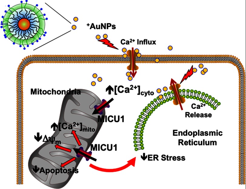
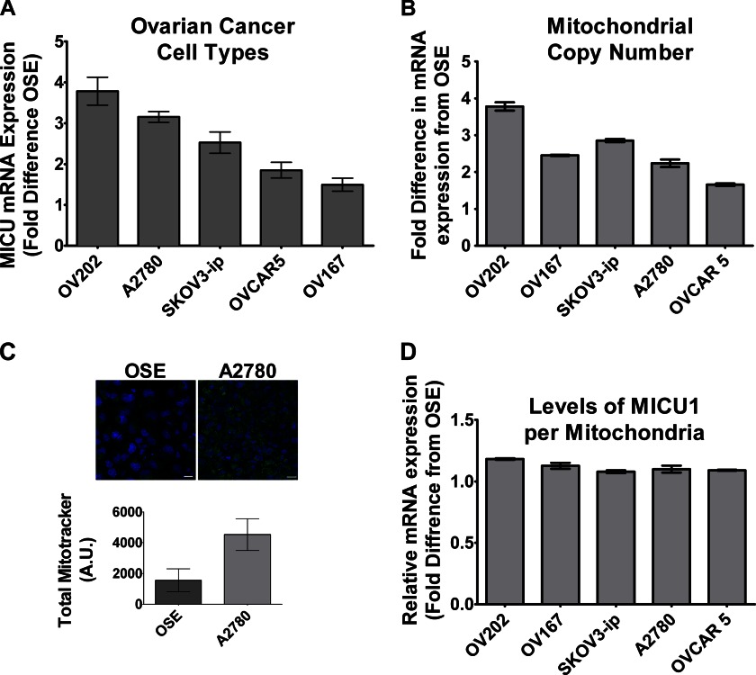
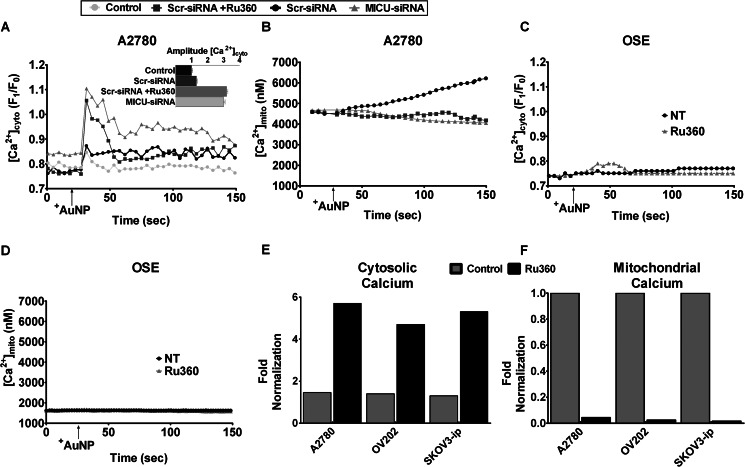
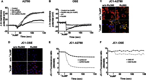
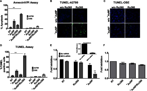
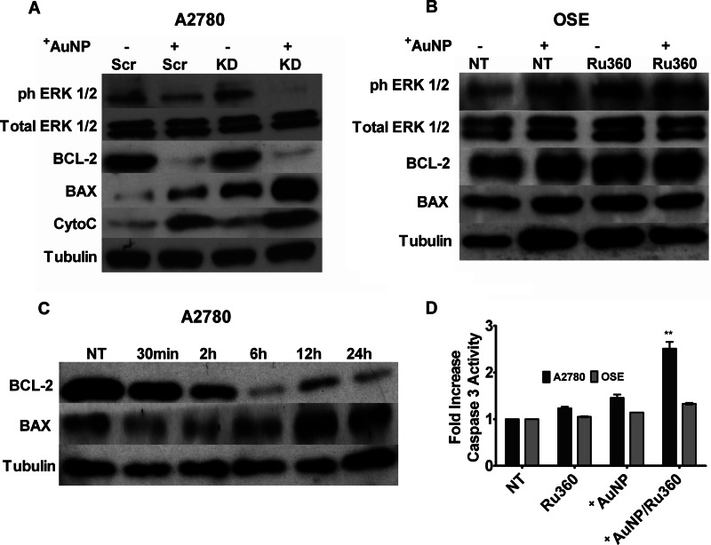
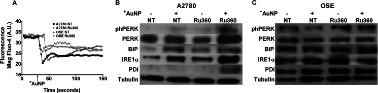
Nanoparticles provide a potent tool for targeting and understanding disease mechanisms. In this regard, cancer cells are surprisingly resistant to the expected toxic effects of positively charged gold nanoparticles ((+)AuNPs). Our investigations led to the identification of MICU1, regulator of mitochondrial calcium uniporter, as a key molecule conferring cancer cells with resistance to (+)AuNPs. The increase in cytosolic [Ca(2+)]cyto in malignant cells induced by (+)AuNPs is counteracted by MICU1, preventing cell death. Pharmacological or siRNA-mediated inhibition of mitochondrial Ca(+2) entry leads to endoplasmic reticulum stress and sensitizes cancer cells to (+)AuNP-induced cytotoxicity. Silencing MICU1 decreases Bcl-2 expression and increases caspase-3 activity and cytosolic cytochrome c levels, thus initiating the mitochondrial pathway for apoptosis: effects further enhanced by (+)AuNPs. This study highlights the potential of nanomaterials as a tool to broaden our understanding of cellular processes, establishes MICU1 as a novel regulator of the machinery in cancer cells that prevents apoptosis, and emphasizes the need to synergize nanoparticle design with understanding of mitochondrial machinery for enhancing targeted cellular toxicity.

Keywords: Apoptosis; Bcl-2; Cancer; Cancer Biology; Caspase; Mitochondrial Apoptosis.


Figures

SCHEME 1.
SCHEME 1.
Role of the mitochondrial uniporter regulator MICU1 in calcium buffering and ER stress. +AuNPs can be used to examine the role of MICU1. +AuNPs cause plasma membrane depolarization, allowing Ca2+ influx, which is normally buffered by mitochondria regulated by MICU1, which also has an antiapoptotic role. Separately, MICU1 may normally prevent ER stress such as that induced by +AuNPs. Thus, we propose that enhanced MICU1 expression or activity is a key mechanism that prevents the desired cytotoxic effects of nanoparticles and other agents in conditions such as cancer.
FIGURE 1.
FIGURE 1.
MICU1 in ovarian cancer cells. A, MICU1 is expressed to a greater extent in different types of ovarian cancer cells as compared with noncancerous controls (OSE) as determined by mRNA measurements. B and C, enhanced MICU1 expression may represent more mitochondria or increased MICU1 per mitochondrion. Real-time PCR of the mitochondrial gene MT-ND1 (Complex I; B) as well as confocal imaging of cells stained with MitoTracker Green (C) show greater number of mitochondria in A2780 malignant cells (blue staining represents DAPI). A. U., absorbance units. D, however, the ratio of MICU1 to MT-ND1 is comparable between malignant and nonmalignant cells. Values are means ± S.D., n = 3.
FIGURE 2.
FIGURE 2.
MICU1-mediated calcium buffering in ovarian cancer cells evaluated using +AuNPs. A and B, in A2780 cells loaded with the fluorescent dyes fura-2 ([Ca2+]cyto) and rhod-2 ([Ca2+]mito), 1 μm +AuNPs caused rapid increase in [Ca2+]cyto (A) but slower sustained increases in [Ca2+]mito (B). Inhibition of uniporter using Ru360 or MICU1 siRNA abrogated [Ca2+]mito responses to +AuNPs, and conversely enhanced [Ca2+]cyto responses. C and D, in contrast to A2780, [Ca2+]cyto and especially [Ca2+]mito responses, or the effects of uniporter inhibition, were substantially smaller in nonmalignant OSE cells. NT, nontreated controls. E and F, the greater role of uniporter in [Ca2+]cyto (E) buffering and [Ca2+]mito (F) responses to +AuNPs is valid in different types of ovarian cancer cells (A2780, OV202, and SKOV3-ip), highlighting the functional importance of MICU1 in malignant cells. Values are means ± S.D., n = 3.
FIGURE 3.
FIGURE 3.
MICU1 modulates ΔΨm responses in malignant cells. A and B, in A2780 cells loaded with the fluorescent ΔΨm indicator DiBASC2(3), +AuNPs caused rapid and substantial membrane depolarization (A), whereas nonmalignant OSE cells showed slight hyperpolarization (B). +AuNP effects on ΔΨm in A2780 cells were enhanced by MICU1 blockade (siRNA). NT, nontreated controls. C–F, these +AuNP effects on ΔΨm were also evident in confocal images of the fluorescent dye JC-1 in malignant cells (C) versus normal cells (D) and corresponding measurements of JC-1 (E and F). Inhibition of uniporter by Ru360 enhanced +AuNP effects on ΔΨm. A similar effect was observed with MICU1 siRNA (not shown). Scale bar = 20 μm; n = 3. w/o +AuNP, without +AuNP.
FIGURE 4.
FIGURE 4.
Uniporter and MICU1 regulate proliferation and apoptosis in malignant cells. A–C, exposure of A2780 cells to +AuNPs induces cellular apoptosis to a certain extent, as shown by annexin V/propidium iodide (PI) assay (A) and TUNEL stain (B and C). NT, nontreated controls. D, the ratio of TUNEL-positive nuclei (green fluorescence) to DAPI (blue fluorescence) was compared and quantified and expressed as the percentage of the total. Ru360 inhibition substantially enhances +AuNP-induced apoptosis (A–D). E, inhibition of MICU1 does not influence base-line cellular proliferation (thymidine incorporation assay) relative to nontreated controls. However, +AuNP-induced cell proliferation is substantially reduced when MICU1 is inhibited (inset shows the efficiency of MICU siRNA knockdown in A2780 cells). F, OSE cells showing a marginal effect under similar conditions. Values are means ± S.D., n = 3. ** indicates significant AuNP effect (p < 0.001). Images shown are representative. Scale bar = 20 μm.
FIGURE 5.
FIGURE 5.
Mechanisms by which MICU1 influences +AuNP-induced apoptosis. A, immunoblots of whole cell lysates from A2780 cells for markers of apoptosis (BAX), antiapoptosis (BCL-2), and proliferation (ERK1/2 and phospho-ERK1/2 (ph-ERK1/2)) showed that +AuNPs increased expression of proapoptotic factors. In the presence of MICU1 siRNA (KD), these effects were substantially enhanced. The enhancement of +AuNP-induced apoptosis by blocking MICU1 (through siRNA; KD) was also demonstrated by increased cytosolic expression of cytochrome c (CytoC). Scr, scrambled siRNA . B, these effects of +AuNPs were absent in OSE cells. NT, nontreated controls. C, the effects in A2780 cells begin after 2 h of exposure to +AuNPs in the knockdown cells. D, separately inhibition by Ru360 leads to increased activity of caspase 3 in A2780 cells with only a modest effect in OSE cells (relative to nontreated controls). Tubulin was used as a loading control. Values are means ± S.D., n = 3. ** indicates significant +AuNP effect (p < 0.001).
FIGURE 6.
FIGURE 6.
Uniporter contributes to ER stress in malignant cells. A, in A2780 cells loaded with the fluorescent ER Ca2+ indicator mag-fluo-4, +AuNPs produced substantial ER Ca2+ release (decreased fluorescence). In the presence of Ru360, base-line ER Ca2+ was higher, but +AuNP-induced release of ER Ca2+ was comparable with control (n = 3). However, ER uptake of Ca2+ was more rapid in Ru360-treated cells. A. U., absorbance units. B and C, these +AuNP-induced changes in ER Ca2+ were paralleled by enhanced expression of ER stress proteins such as phospho-PERK (phPERK), total PERK, BiP, and IRE1α (but not protein disulfide isomerase) in A2780 cells (B) but not in OSE cells (C). NT, nontreated controls.

Similar articles

Cited by

References

    1. Arvizo R. R., Rana S., Miranda O. R., Bhattacharya R., Rotello V. M., Mukherjee P. (2011) Mechanism of anti-angiogenic property of gold nanoparticles: role of nanoparticle size and surface charge. Nanomedicine 7, 580–587 - PMC - PubMed
    1. Bhattacharyya S., Singh R. D., Pagano R., Robertson J. D., Bhattacharya R., Mukherjee P. (2012) Switching the targeting pathways of a therapeutic antibody by nanodesign. Angew. Chem. Int. Ed. Engl. 51, 1563–1567 - PMC - PubMed
    1. Bhirde A. A., Patel V., Gavard J., Zhang G., Sousa A. A., Masedunskas A., Leapman R. D., Weigert R., Gutkind J. S., Rusling J. F. (2009) Targeted killing of cancer cells in vivo and in vitro with EGF-directed carbon nanotube-based drug delivery. ACS Nano 3, 307–316 - PMC - PubMed
    1. Agasti S. S., Chompoosor A., You C.-C., Ghosh P., Kim C. K., Rotello V. M. (2009) Photoregulated release of caged anticancer drugs from gold nanoparticles. J. Am. Chem. Soc. 131, 5728–5729 - PMC - PubMed
    1. Zhu Z. J., Ghosh P. S., Miranda O. R., Vachet R. W., Rotello V. M. (2008) Multiplexed screening of cellular uptake of gold nanoparticles using laser desorption/ionization mass spectrometry. J. Am. Chem. Soc. 130, 14139–14143 - PMC - PubMed

Publication types

MeSH terms