. 2013 Jun 7;288(23):16403-16415.
doi: 10.1074/jbc.M113.474593. Epub 2013 Apr 24.

Phosphorylation of serine 1137/1138 of mouse insulin receptor substrate (IRS) 2 regulates cAMP-dependent binding to 14-3-3 proteins and IRS2 protein degradation

Affiliations

Phosphorylation of serine 1137/1138 of mouse insulin receptor substrate (IRS) 2 regulates cAMP-dependent binding to 14-3-3 proteins and IRS2 protein degradation

Sabine S Neukamm et al. J Biol Chem. .

Abstract

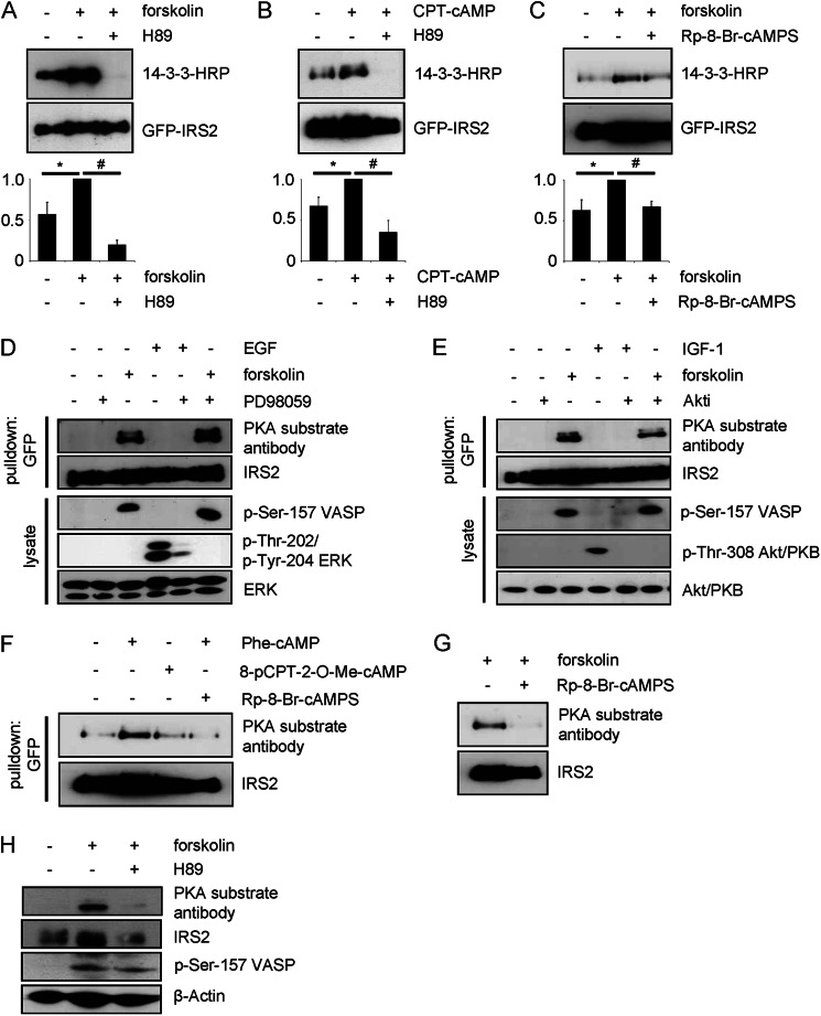
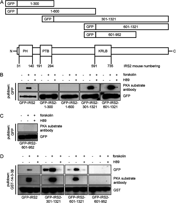
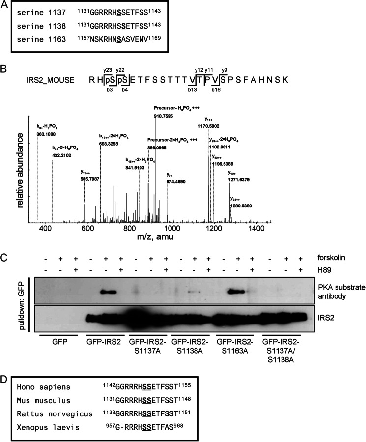
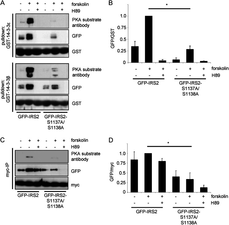
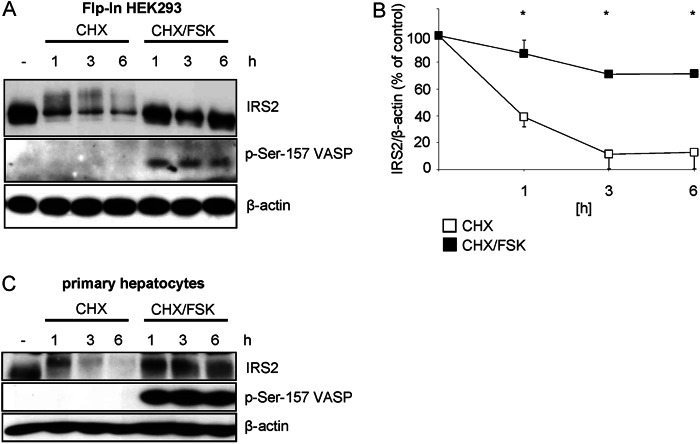
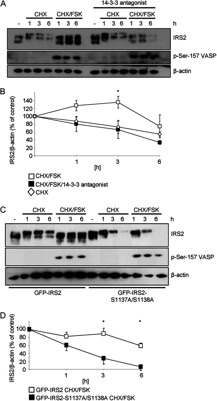
Insulin receptor substrate (IRS) 2 as intermediate docking platform transduces the insulin/IGF-1 (insulin like growth factor 1) signal to intracellular effector molecules that regulate glucose homeostasis, β-cell growth, and survival. Previously, IRS2 has been identified as a 14-3-3 interaction protein. 14-3-3 proteins can bind their target proteins via phosphorylated serine/threonine residues located within distinct motifs. In this study the binding of 14-3-3 to IRS2 upon stimulation with forskolin or the cAMP analog 8-(4-chlorophenylthio)-cAMP was demonstrated in HEK293 cells. Binding was reduced with PKA inhibitors H89 or Rp-8-Br-cAMPS. Phosphorylation of IRS2 on PKA consensus motifs was induced by forskolin and the PKA activator N(6)-Phe-cAMP and prevented by both PKA inhibitors. The amino acid region after position 952 on IRS2 was identified as the 14-3-3 binding region by GST-14-3-3 pulldown assays. Mass spectrometric analysis revealed serine 1137 and serine 1138 as cAMP-dependent, potential PKA phosphorylation sites. Mutation of serine 1137/1138 to alanine strongly reduced the cAMP-dependent 14-3-3 binding. Application of cycloheximide revealed that forskolin enhanced IRS2 protein stability in HEK293 cells stably expressing IRS2 as well as in primary hepatocytes. Stimulation with forskolin did not increase protein stability either in the presence of a 14-3-3 antagonist or in the double 1137/1138 alanine mutant. Thus the reduced IRS2 protein degradation was dependent on the interaction with 14-3-3 proteins and the presence of serine 1137/1138. We present serine 1137/1138 as novel cAMP-dependent phosphorylation sites on IRS2 and show their importance in 14-3-3 binding and IRS2 protein stability.

Keywords: 14-3-3; Cyclic AMP (cAMP); IRS2; Insulin; Phosphorylation; Protein Degradation; Protein Kinase A (PKA).


Figures

FIGURE 1.
FIGURE 1.
14-3-3 interacts with IRS2 upon elevated cAMP levels and PKA phosphorylates IRS2. A, HEK293 cells were transiently transfected with GFP-IRS2. Cells were incubated for 30 min with 20 μm forskolin alone or after preincubation with 30 μm H89 for 30 min. 100 μg of protein was used for GFP pulldown. 14-3-3 binding was visualized by performing an overlay assay and the membrane was stripped and reprobed for GFP-IRS2 as expression and loading control. Densitometric analysis of 14-3-3 binding is shown as band intensities normalized for GFP-IRS2. GFP-IRS2 treated with forskolin was set as 1 (mean ± S.E., n = 3, *, p < 0.05, forskolin versus unstimulated; #, <0.05, forskolin versus H89). B, cells were stimulated with 100 μm CPT-cAMP for 30 min or after preincubation with 30 μm H89 for 30 min. 200 μg of protein was used for GFP pulldown. GFP-IRS2 treated with CPT-cAMP was set as 1 (mean ± S.E., n = 3, *, p < 0.05, CPT-cAMP versus unstimulated; #, <0.05, CPT-cAMP versus H89). C, cells were incubated for 30 min with 20 μm forskolin alone or after preincubation with 100 μm Rp-8-Br-cAMPS for 30 min. 400 μg of protein was used for GFP pulldown. GFP-IRS2 treated with forskolin was set as 1 (mean ± S.E., n = 3, *, p < 0.05, forskolin versus unstimulated; #, <0.05, forskolin versus Rp-8-Br-cAMPS). D and E, GFP-IRS2 was transiently expressed in HEK293 cells and cells were incubated for 30 min with 1 μm Akt inhibitor VIII Akti 1/2 (Akti), 20 μm forskolin, 50 ng/ml of IGF-1, 50 μm PD98059, and 100 ng/ml of EGF as indicated. 200 μg of total protein was used for GFP pulldown. Membrane was incubated with PKA substrate antibody. Phosphorylation of serine 157 of VASP was assessed as forskolin stimulation control, phosphorylation of threonine 308 of Akt/PKB as IGF-1 stimulation control, whereas phosphorylation of threonine 202/tyrosine 204 of ERK was checked as control for successful EGF stimulation. Corresponding IRS2, Akt/PKB, and ERK reblots are also shown (n = 3). F and G, GFP-IRS2 was expressed transiently in HEK293 cells. Cells were incubated with the following reagents for 30 min: 100 μm N6-Phe-cAMP, 100 μm 8-pCPT-2′-O-Me-cAMP, 100 μm Rp-8-Br-cAMPS or 20 μm forskolin. 400 μg of protein was used for GFP pulldown. Membrane was incubated with PKA substrate antibody, after which it was stripped and reprobed with IRS2 antibody (n = 3). H, primary hepatocytes were incubated with 20 μm forskolin alone or after preincubation with 30 μm H89 for 30 min, respectively. 100 μg of protein was separated on SDS gel and membranes were incubated with PKA substrate antibody, phosphoserine 157 VASP antibody, or β-actin antibody. PKA substrate antibody membrane was stripped and reprobed with IRS2 antibody (n = 3).
FIGURE 2.
FIGURE 2.
Site(s) for PKA phosphorylation on IRS2 are located after amino acid position 952. A, schematic illustration of IRS2 and IRS2 fragments. Five fragments spanning different regions of the IRS2 protein were generated according to mouse numbering: GFP-IRS2-(1–300), GFP-IRS2-(1–600), GFP-IRS2-(301–1321), GFP-IRS2-(601–1321), and GFP-IRS2-(601–952). All constructs were GFP tagged at the N terminus. B and C, HEK293 cells were transiently transfected with GFP-IRS2, GFP-IRS2-(1–300), GFP-IRS2-(1–600), GFP-IRS2-(301–1321), GFP-IRS2-(601–1321), or GFP-IRS2-(601–952). Cells were incubated for 30 min with 20 μm forskolin alone or after preincubation with 30 μm H89 for 30 min. 100–200 μg of protein was used for GFP pulldown and after separation on 5–15% SDS gels and Western blotting, the membrane was incubated with PKA substrate antibody. Stripping and reprobing with GFP antibody as expression and loading control is also shown (n = 3). D, GFP-IRS2, GFP-IRS2-(301–1321), GFP-IRS2-(601–1321), and GFP-IRS2-(601–952) were transiently expressed in HEK293 cells. Stimulation was carried out with 20 μm forskolin for 30 min or 30 μm H89 for 30 min prior forskolin stimulation. Lysate containing 250 μg of total protein was incubated with 2 μg of GST-14-3-3β for 2 h and samples were separated by SDS-PAGE. Transfer onto nitrocellulose membranes followed by incubation with GFP antibody to visualize the amount of protein that interacted with GST-14-3-3 and with GST to ensure equal pulldown and loading. Incubation with PKA substrate antibody was carried out to visualize phosphorylation of PKA motifs (n = 3).
FIGURE 3.
FIGURE 3.
Mass spectrometry identifies three candidate residues in IRS2 for phosphorylation by PKA. A, sequences surrounding the potential PKA phosphorylation sites serine 1137, serine 1138, and serine 1163 are shown according to IRS2 mouse numbering. B, MS/MS analysis of a tryptic peptide with the parent mass (M+3H) = 951.4145 revealed a mass spectrum consisting of single- and double-charged fragment masses (preferably ions most relevant to phosphorylation sites are annotated) matching the doubly phosphorylated IRS2-peptide sequence RHpSpSETFSSTTTVTPVSPSFAHNSK. C, HEK293 cells were transiently transfected with GFP, GFP-IRS2, GFP-IRS2-S1137A, GFP-IRS2-S1138A, GFP-IRS2-S1163A, or GFP-IRS2-S1137A/S1138A. Cells were stimulated with 20 μm forskolin for 30 min alone or after pretreatment with 30 μm H89 for 30 min. GFP pulldown assay was performed with 200 μg of total protein and after SDS-PAGE and Western blotting the membrane was incubated with PKA substrate antibody. The corresponding blot reprobed with IRS2 antibody is also shown (n = 3). D, amino acid areas surrounding serine 1137 and serine 1138 from IRS2 in different species are shown: Homo sapiens (NP_003740.2), Mus musculus (NP_001074681.1), Rattus norvegicus (NP_001162104.1), and Xenopus laevis (AAH72768.1).
FIGURE 4.
FIGURE 4.
Mutation of serine 1137/1138 of IRS2 significantly reduces interaction with 14-3-3. A, GFP-IRS2 and GFP-IRS2-S1137A/S1138A were transiently expressed in HEK293 cells and stimulated for 30 min with 20 μm forskolin alone or after preincubation with 30 μm H89 for 30 min. Lysates containing 250 μg of total protein were incubated with 2 μg of GST-14-3-3ϵ or GST-14-3-3β for 2 h, after which they were separated by SDS-PAGE. Transfer onto nitrocellulose membranes was followed by incubation with GFP antibody to visualize the amount of protein that interacted with GST-14-3-3, with PKA substrate antibody and with GST antibody to ensure equal pulldown and loading. B, densitometric analysis of pulldown with GST-14-3-3ϵ. GFP signal intensities were normalized to GST and GFP-IRS2 stimulated with forskolin was set as 1 (mean ± S.E., n = 3, *, p < 0.05 GFP-IRS2 forskolin versus GFP-IRS2-S1137A/S1138A forskolin-treated. C, HEK293 cells were transiently co-transfected with GFP-IRS2 and Myc-14-3-3γ or GFP-IRS2-S1137A/S1138A and Myc-14-3-3γ, and incubated for 30 min with 20 μm forskolin alone or after pretreatment with 30 μm H89 for 30 min. Lysates containing 200 μg of protein were incubated with Myc antibody and Protein G-Sepharose for 3 h at 4 °C. Samples were separated on 5–15% SDS gels and after Western blotting, the membranes were incubated with GFP antibody, PKA substrate antibody and Myc antibody. D, densitometric analysis of co-immunoprecipitated GFP. Band intensities were normalized for Myc and GFP-IRS2 treated with forskolin was set as 1 (mean ± S.E., n = 3, *, p < 0.05, GFP-IRS2 forskolin versus GFP-IRS2-S1137A/S1138A forskolin-treated).
FIGURE 5.
FIGURE 5.
Forskolin prevents protein degradation of IRS2. A, Flp-In HEK293 cells stably expressing IRS2 were incubated with 25 μg/ml of cycloheximide (CHX) alone or in combination with 20 μm forskolin (FSK) for 1, 3, and 6 h. 10 μg of protein was separated and membranes were incubated with antibodies against IRS2, phosphorylated serine 157 of VASP as stimulation control and β-actin as loading control. B, densitometric analysis of IRS2 protein bands. IRS2 content was normalized for β-actin and IRS2 from untreated cells was set as 100% (mean ± S.E., n = 8, *, p < 0.05 cycloheximide (open squares) versus cycloheximide/forskolin-treated (filled squares)). C, hepatocytes were isolated from male C57Bl/6 mice, plated onto 6-well plates, treated as described in A and 100 μg of protein was analyzed (n = 3).
FIGURE 6.
FIGURE 6.
14-3-3 proteins and serine 1137/1138 are important for IRS2 protein stability. A, Flp-In HEK293 cells stably expressing GFP-IRS2 were treated for the indicated time points with 25 μg/ml of cycloheximide (CHX) alone or in combination with 20 μm forskolin (FSK). Another set of cells was treated with 20 μm 14-3-3 antagonist overnight and during the incubation times. 25 μg of protein was separated by SDS-PAGE and membranes were incubated with following antibodies: IRS2, phosphoserine 157 of VASP and β-actin. B, densitometric analysis of IRS2 protein degradation. IRS2 content was normalized for β-actin and IRS2 from untreated cells was set as 100% (mean ± S.E., n = 4, *, p < 0.05 cycloheximide/forskolin (open squares) versus cycloheximide/forskolin/14-3-3 antagonist-treated (filled squares) at 3 h). C, Flp-In HEK293 cells stably expressing GFP-IRS2 or GFP-IRS2-S1137A/S1138A were incubated with 25 μg/ml of CHX and 20 μm FSK for 1, 3, and 6 h. 25 μg of protein was separated by SDS-PAGE and membranes were incubated with IRS2 antibody, phosphoserine 157 of VASP and β-actin antibody. D, the extent of IRS2 protein degradation was quantified by scanning immunoblots and normalization of IRS2 signal intensities for β-actin. Untreated cells expressing GFP-IRS2 or GFP-IRS2-S1137A/S1138A were set to 100% (mean ± S.E., n = 4, *, p < 0.05, GFP-IRS2 (open squares) versus GFP-IRS2-S1137A/S1138A (filled squares) at 3 and 6 h).

Similar articles

Cited by

References

    1. Kubota N., Tobe K., Terauchi Y., Eto K., Yamauchi T., Suzuki R., Tsubamoto Y., Komeda K., Nakano R., Miki H., Satoh S., Sekihara H., Sciacchitano S., Lesniak M., Aizawa S., Nagai R., Kimura S., Akanuma Y., Taylor S. I., Kadowaki T. (2000) Disruption of insulin receptor substrate 2 causes type 2 diabetes because of liver insulin resistance and lack of compensatory beta-cell hyperplasia. Diabetes 49, 1880–1889 - PubMed
    1. Kubota N., Kubota T., Itoh S., Kumagai H., Kozono H., Takamoto I., Mineyama T., Ogata H., Tokuyama K., Ohsugi M., Sasako T., Moroi M., Sugi K., Kakuta S., Iwakura Y., Noda T., Ohnishi S., Nagai R., Tobe K., Terauchi Y., Ueki K., Kadowaki T. (2008) Dynamic functional relay between insulin receptor substrate 1 and 2 in hepatic insulin signaling during fasting and feeding. Cell Metab. 8, 49–64 - PubMed
    1. Withers D. J., Gutierrez J. S., Towery H., Burks D. J., Ren J. M., Previs S., Zhang Y., Bernal D., Pons S., Shulman G. I., Bonner-Weir S., White M. F. (1998) Disruption of IRS-2 causes type 2 diabetes in mice. Nature 391, 900–904 - PubMed
    1. Schuppin G. T., Pons S., Hügl S., Aiello L. P., King G. L., White M., Rhodes C. J. (1998) A specific increased expression of insulin receptor substrate 2 in pancreatic beta-cell lines is involved in mediating serum-stimulated beta-cell growth. Diabetes 47, 1074–1085 - PubMed
    1. Hennige A. M., Burks D. J., Ozcan U., Kulkarni R. N., Ye J., Park S., Schubert M., Fisher T. L., Dow M. A., Leshan R., Zakaria M., Mossa-Basha M., White M. F. (2003) Up-regulation of insulin receptor substrate-2 in pancreatic beta cells prevents diabetes. J. Clin. Invest. 112, 1521–1532 - PMC - PubMed

Publication types

MeSH terms

LinkOut - more resources