. 2013 May 31;288(22):15437-41.
doi: 10.1074/jbc.C112.446682. Epub 2013 Apr 24.

Overexpression of USP14 protease reduces I-κB protein levels and increases cytokine release in lung epithelial cells

Affiliations

Overexpression of USP14 protease reduces I-κB protein levels and increases cytokine release in lung epithelial cells

Rachel K Mialki et al. J Biol Chem. .

Abstract

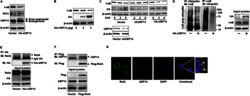
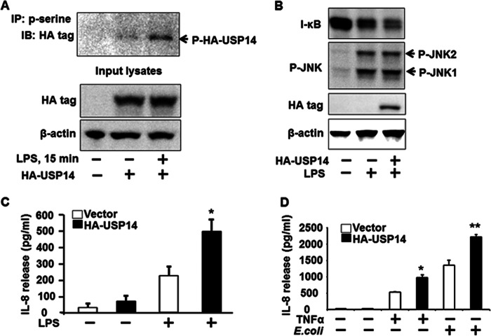
The ubiquitin-proteasome system is the major pathway of non-lysosomal intracellular protein degradation, playing an important role in a variety of cellular responses including cell division, proliferation, and apoptosis. Ubiquitin-specific protease 14 (USP14) is a component of proteasome regulatory subunit 19 S that regulates deubiquitinated proteins entering inside the proteasome core 20 S. The role of USP14 in protein degradation is still controversial. Several studies suggest that USP14 plays an inhibitory role in protein degradation. Here, in contrast, overexpression of USP14 induced I-κB degradation, which increased cytokine release in lung epithelial cells. Overexpression of HA-tagged USP14 (HA-USP14) reduced I-κB protein levels by increasing the I-κB degradation rate in mouse lung epithelial cells (MLE12). I-κB polyubiquitination was reduced in HA-USP14-overexpressed MLE12 cells, suggesting that USP14 regulates I-κB degradation by removing its ubiquitin chain, thus promoting the deubiquitinated I-κB degradation within the proteasome. Interestingly, we found that USP14 was associated with RelA, a binding partner of I-κB, suggesting that RelA is the linker between USP14 and I-κB. Lipopolysaccharide (LPS) treatment induced serine phosphorylation of USP14 as well as further reducing I-κB levels in HA-USP14-overexpressed MLE12 cells as compared with empty vector transfected cells. Further, overexpression of HA-USP14 increased the LPS-, TNFα-, or Escherichia coli-induced IL-8 release in human lung epithelial cells. This study suggests that USP14 removes the ubiquitin chain of I-κB, therefore inducing I-κB degradation and increasing cytokine release in lung epithelial cells.

Keywords: Cytokine; Deubiquitination; NF-κB Transcription Factor; Protein Phosphorylation; Protein degradation; USP14.


Figures

FIGURE 1.
FIGURE 1.
Overexpression of USP14 reduces I-κB levels and ubiquitination via association with RelA. A, MLE12 cells were transfected with an HA-USP14 plasmid (2 μg) for 48 h. Cell lysates were subjected to immunoblotting with antibodies to I-κB, IKKα, USP14, and β-actin. B, MLE12 cells were transfected with an HA-USP14 plasmid (2 and 4 μg) for 48 h. Cell lysates were subjected to I-κB, HA tag, and β-actin. C, MLE12 cells were transfected with an empty vector or an HA-USP14 plasmid for 48 h or with USP14 shRNA (shUSP14) for 72 h. Cell lysates were subjected to immunoblotting with antibodies to I-κB, HA tag, and β-actin (upper panel). Cell lysates from vector or shUSP14 transfected cells were analyzed with USP14 and β-actin antibodies (lower panel). CHX, cycloheximide. D, MLE12 cells were transfected with an empty vector or an HA-USP14 plasmid (3 μg) for 48 h prior to MG-132 (20 μm, 4 h) treatment. Cell lysates were subjected to immunoprecipitation (IP) with a ubiquitin antibody followed by I-κB immunoblotting (IB, left panel). Cell lysates were subjected to immunoprecipitation with an I-κB ubiquitin antibody followed by ubiquitin immunoblotting (IB, middle panel). Input lysates were subjected to immunoblotting with antibodies to HA tag, I-κB, and β-actin (right panel). E, MLE12 cells were transfected with an empty vector or an HA-USP14 plasmid (3 μg) for 48 h. Cell lysates were subjected to immunoprecipitation with an HA tag antibody followed by immunoblottings with antibodies to RelA and HA tag. Input lysates were subjected to immunoblotting with antibodies to RelA, HA tag, and β-actin. F, MLE12 cells were transfected with an empty vector or a FLAG-RelA plasmid (3 μg) for 48 h. Cell lysates were subjected to immunoprecipitation with a FLAG tag antibody followed by immunoblottings with antibodies to USP14 and FLAG tag. Input lysates were subjected to immunoblotting with antibodies to USP14, FLAG tag, and β-actin. G, MLE12 cells grown on glass bottom dishes were fixed and immunostained with antibodies to RelA (shown in green) and USP14 (shown in red). Nuclei were stained with DAPI (blue). Shown are representative images from three independent experiments.
FIGURE 2.
FIGURE 2.
Overexpression of USP14 promotes LPS-induced IL-8 release. A, MLE12 cells were transfected with an empty vector or an HA-USP14 plasmid (3 μg) for 48 h. Cell lysates were subjected to immunoprecipitation (IP) with a phospho-serine (p-serine) antibody followed by immunoblottings (IB) with an antibody to HA tag. Input lysates were subjected to immunoblotting with antibodies to HA tag and β-actin. B, MLE12 cells were transfected with an empty vector or an HA-USP14 plasmid (3 μg) for 48 h prior to LPS treatment (10 μg/ml, 30 min). Cell lysates were subjected to immunoblotting with antibodies to I-κB, phospho-JNK (p-JNK), HA tag, and β-actin. C, Beas2B cells were transfected with an empty vector or an HA-USP14 plasmid (3 μg) for 48 h prior to LPS treatment (10 μg/ml, 3 h). Media were collected, and IL-8 release was measured by IL-8 ELISA kit. *, p < 0.01 as compared with cells treated with LPS alone. D, Beas2B cells were transfected with an empty vector or an HA-USP14 plasmid (3 μg) for 48 h prior to TNFα (10 μg/ml) treatment or E. coli infection (10 multiplicity of infection) for 3 h. Media were collected, and IL-8 release was measured by IL-8 ELISA kit. *, p < 0.01 as compared with cells treated with TNFα alone; **, p < 0.01 as compared with cells treated with E. coli alone.

Similar articles

Cited by

References

    1. Hershko A., Ciechanover A. (1992) The ubiquitin system for protein degradation. Annu. Rev. Biochem. 61, 761–807 - PubMed
    1. Schwartz A. L., Ciechanover A. (1992) Ubiquitin-mediated protein modification and degradation. Am. J. Respir. Cell Mol. Biol. 7, 463–468 - PubMed
    1. Ugai S., Tamura T., Tanahashi N., Takai S., Komi N., Chung C. H., Tanaka K., Ichihara A. (1993) Purification and characterization of the 26S proteasome complex catalyzing ATP-dependent breakdown of ubiquitin-ligated proteins from rat liver. J. Biochem. 113, 754–768 - PubMed
    1. Saeki Y., Tanaka K. (2012) Assembly and function of the proteasome. Methods Mol. Biol. 832, 315–337 - PubMed
    1. Kim H. M., Yu Y., Cheng Y. (2011) Structure characterization of the 26S proteasome. Biochim. Biophys. Acta 1809, 67–79 - PMC - PubMed

Publication types

MeSH terms

LinkOut - more resources