. 2013 Jul;43(7):1896-906.
doi: 10.1002/eji.201242792. Epub 2013 May 28.

IRF-5 and NF-κB p50 co-regulate IFN-β and IL-6 expression in TLR9-stimulated human plasmacytoid dendritic cells

Affiliations

IRF-5 and NF-κB p50 co-regulate IFN-β and IL-6 expression in TLR9-stimulated human plasmacytoid dendritic cells

Folkert Steinhagen et al. Eur J Immunol. 2013 Jul.

Abstract

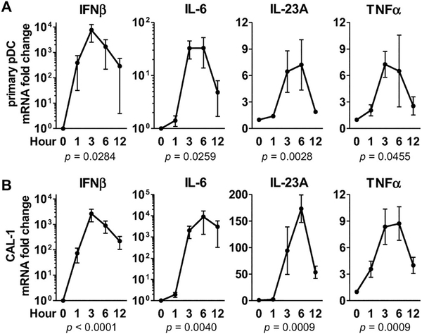
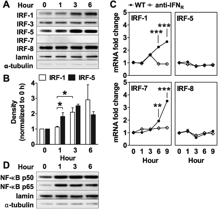
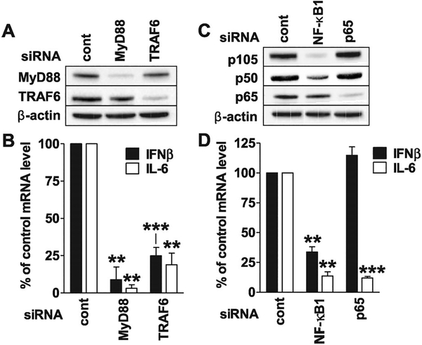
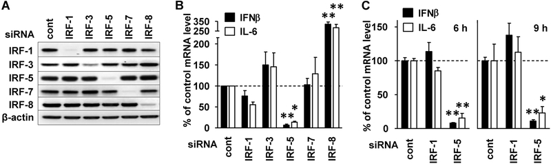
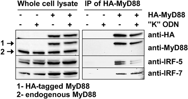
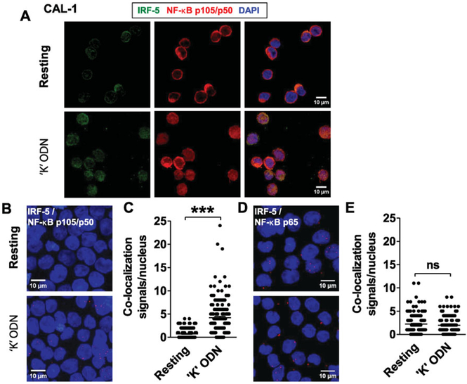
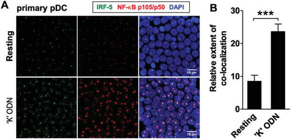
Synthetic oligonucleotides (ODN) expressing CpG motifs mimic the ability of bacterial DNA to trigger the innate immune system via TLR9. Plasmacytoid dendritic cells (pDCs) make a critical contribution to the ensuing immune response. This work examines the induction of antiviral (IFN-β) and pro-inflammatory (IL-6) cytokines by CpG-stimulated human pDCs and the human CAL-1 pDC cell line. Results show that interferon regulatory factor-5 (IRF-5) and NF-κB p50 are key co-regulators of IFN-β and IL-6 expression following TLR9-mediated activation of human pDCs. The nuclear accumulation of IRF-1 was also observed, but this was a late event that was dependant on type 1 IFN and unrelated to the initiation of gene expression. IRF-8 was identified as a novel negative regulator of gene activation in CpG-stimulated pDCs. As variants of IRF-5 and IRF-8 were recently found to correlate with susceptibility to certain autoimmune diseases, these findings are relevant to our understanding of the pharmacologic effects of "K" ODN and the role of TLR9 ligation under physiologic, pathologic, and therapeutic conditions.

Keywords: CpG oligonucleotide; Dendritic cell; IRF-5; NF-κB; TLR9.


Conflict of interest statement

Conflict of interest:

Dr. Dennis Klinman and members of his lab are co-inventors on a number of patents concerning CpG ODN and their use. All rights to these patents have been assigned to the Federal government.

Figures

Figure 1.
Figure 1.
Cytokine production by primary human pDCs and CAL-1 cells stimulated with “K” ODN. CAL-1 cells and freshly isolated human pDCs were stimulated with 1 μM of “K” ODN for the indicated times. mRNA levels for IFN-β, IL-6, IL-23A and TNF-α were assessed by RT-PCR using GAPDH as an endogenous control. Data represent the mean ± SEM from — three to four independent experiments. Overall statistical significance between means was determined using a repeated measures ANOVA.
Figure 2.
Figure 2.
Nuclear translocation of IRF and NF-κB transcription factors and induction of IRF expression following “K” ODN stimulation. CAL-1 cells were incubated with 1 μM of “K” ODN for the indicated times. Nuclear lysates were extracted and analyzed by immunoblotting for changes in the concentration of (A) various IRFs and (D) NF-κB p50 and p65. (B) The fold increase in nuclear levels of IRF-1 and IRF-5 relative to untreated controls was determined by densitometric analysis. Lamin and α-tubulin were used as quality controls to assess nuclear extract purity and loading levels. (C) The effect of adding anti-type 1 IFN receptor antibody (anti-IFNR) to “K” ODN stimulated cultures was examined. Data represent the mean + SEM from three independent experiments. *p < 0.05, **p < 0.01, ***p < 0.001, Student’s t-test.
Figure 3.
Figure 3.
MyD88/TRAF6 and NF-κB1 and NF-κB p65 influence “K” ODN mediated gene activation. CAL-1 cells were transfected with siRNA to knockdown gene expression. Whole cells lysates were analyzed by immunoblot to evaluate the efficiency of (A) MyD88 and TRAF6 or (C) NF-κB1 (p105/p50) and NF-κB p65 protein knockdown. β-actin was used as a loading control. (B and D) siRNA-transfected CAL-1 cells were stimulated with 1 μM of “K” ODN for 3 h. The percent reduction in IL-6 and IFN-β mRNA levels were assessed by RT-PCR with GAPDH as an endogenous control. Changes in mRNA levels were evaluated by comparison to identically stimulated cells transfected with control siRNA in each experiment. Data represent the mean + SEM from three independent experiments. **p < 0.01, ***p < 0.001 (one-way ANOVA).
Figure 4.
Figure 4.
Influence of IRFs on “K” ODN mediated gene activation. CAL-1 cells were transfected with various IRF siRNAs to knockdown gene expression. (A) Whole cell lysates were analyzed by immunoblot to evaluate the efficiency of protein knockdown. β-actin was used as a loading control. (B) siRNA-transfected CAL-1 cells were stimulated with 1 μM of “K” ODN for 3 h. (C) The effect of IRF-1 and IRF-5 knockdown was further evaluated at 6 and 9 h. The fold change in IFN-β and IL-6 mRNA levels was assessed by RT-PCR with GAPDH as an endogenous control. Changes in mRNA level were evaluated by comparison to unstimulated cells transfected with control siRNA in each experiment. Data represent the mean + SEM from three independent experiments. *p < 0.05, **p < 0.01 (one-way ANOVA).
Figure 5.
Figure 5.
Association of IRF-5 with MyD88. CAL-1 cells were transfected with a plasmid encoding HA-tagged MyD88 and then stimulated for 30 min with 1 μM of “K” ODN. A whole cell lysate (left panel) was prepared and analyzed by immunoblot for the presence of MyD88 (endogenous or HAtagged), IRF-5, and IRF-7. The right panel shows proteins that co-precipitated with the HA-tagged MyD88. Data are representative of results obtained in two independent experiments.
Figure 6.
Figure 6.
Co-localization of IRF-5 with NF-κB p50 in CpG-stimulated CAL-1 cells. (A) CAL-1 cells were stimulated for 60 min with 1 μM of “K” ODN. Cells were fixed, permeabilized, and stained to detect IRF-5 (green) and NF-κB p105/p50 (red). Nuclei were stained with DAPI (blue). (B–E) CAL-1 cells were stimulated for 30 min with 1 μM of “K” ODN. The co-localization (red dots) of (B) IRF-5 with NF-κB p105/p50 and (D) IRF-5 with p65 was determined by proximity ligation assay. The frequency of IRF-5/NF-κB p50 (C) and IRF-5/p65 (D) co-localization events per CAL-1 nucleus is shown (N > 150 cells per treatment from two independent experiments). Scale bars: 10 μM. ***p < 0.0001, Student’s t-test.
Figure 7.
Figure 7.
Co-localization of IRF-5 with NF-κB p105/p50 in CpG-stimulated primary pDCs. (A) Primary pDCs were stimulated for 60 min with 1 μM of “K” ODN. Cells were fixed onto slides, permeabilized, and stained to detect IRF-5 (green), NF-κB p105/p50 (red), and nuclei (blue) as described in Figure 5. (B) Four areas per slide were analyzed using Image J software to detect and quantify IRF-5/NF-κB p50 nuclear co-localization signals. Results represent the relative number of co-localization events per treatment. Data are shown as mean +SD and are representative of one of three independent experiments performed with similar results. Scale bars: 10 μM. ***p < 0.0001, Student’s t-test.

Comment in

References

    1. Kawai T and Akira S, The role of pattern-recognition receptors in innate immunity: update on Toll-like receptors. Nat. Immunol 2010. 11: 373–384. - PubMed
    1. Jarrossay D, Napolitani G, Colonna M, Sallusto F and Lanzavecchia A, Specialization and complementarity in microbial molecule recognition by human myeloid and plasmacytoid dendritic cells. Eur. J. Immunol 2001. 31: 3388–3393. - PubMed
    1. Kadowaki N, Ho S, Antonenko S, Malefyt RW, Kastelein RA, Bazan F and Liu YJ, Subsets of human dendritic cell precursors express different Toll-like receptors and respond to different microbial antigens. J. Exp. Med 2001. 194: 863–869. - PMC - PubMed
    1. Haining WN, Davies J, Kanzler H, Drury L, Brenn T, Evans J, Angelosanto J et al., CpG oligodeoxynucleotides alter lymphocyte and dendritic cell trafficking in humans. Clin. Cancer Res. 2008. 14: 5626–5634. - PubMed
    1. Steinhagen F, Kinjo T, Bode C and Klinman DM, TLR-based immune adjuvants. Vaccine 2011. 29: 3341–3355. - PMC - PubMed

Publication types

MeSH terms