. 2013 Apr 24;33(17):7548-58.
doi: 10.1523/JNEUROSCI.2764-12.2013.

Homeostatic responses fail to correct defective amygdala inhibitory circuit maturation in fragile X syndrome

Affiliations

Homeostatic responses fail to correct defective amygdala inhibitory circuit maturation in fragile X syndrome

Rebecca L Vislay et al. J Neurosci. .

Abstract

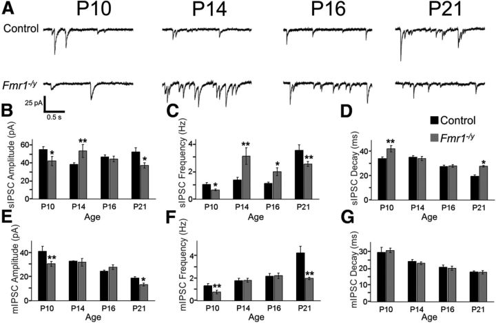
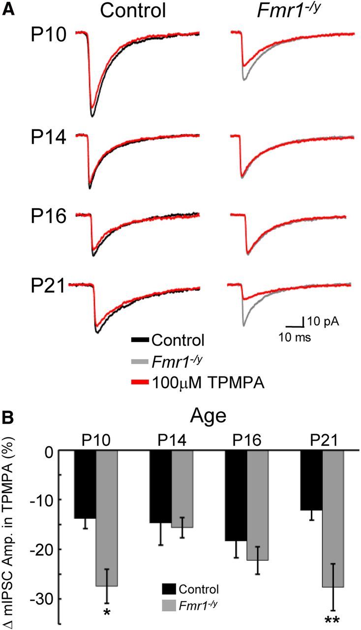
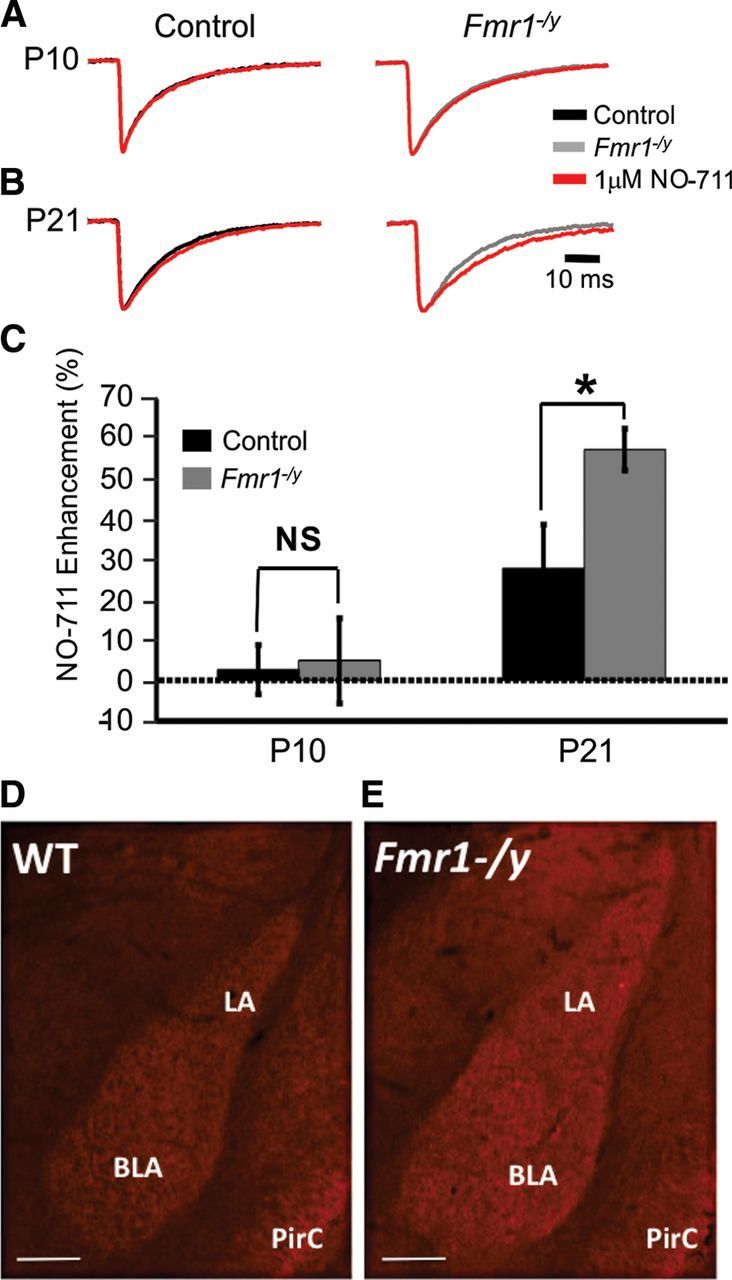
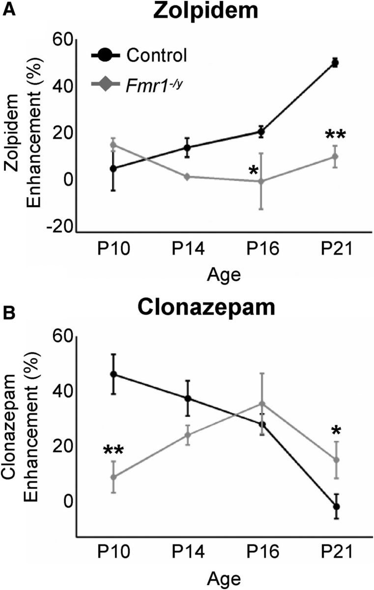
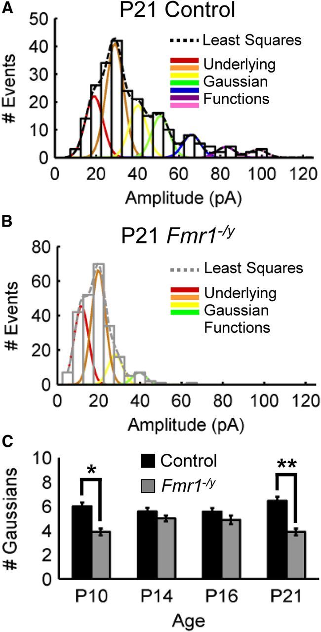
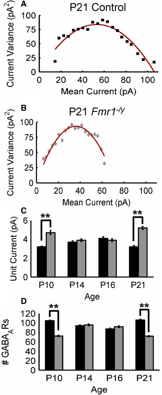
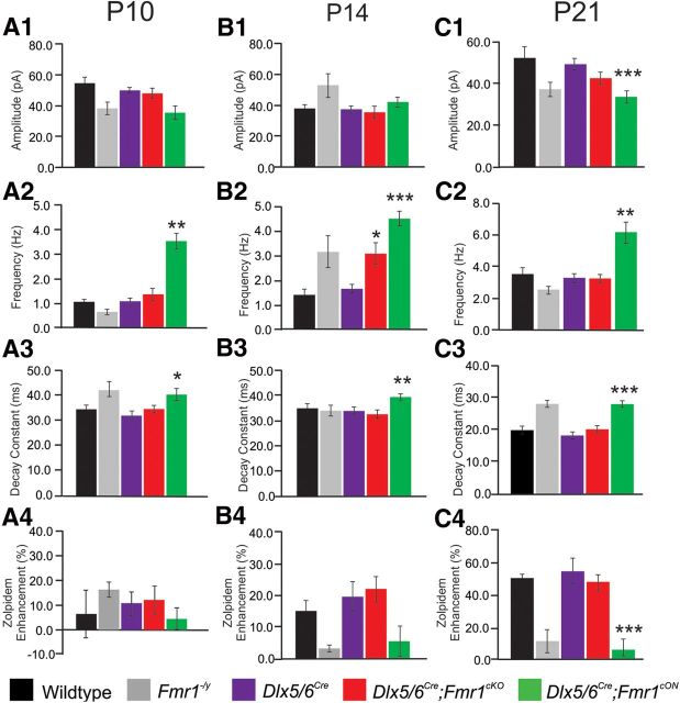
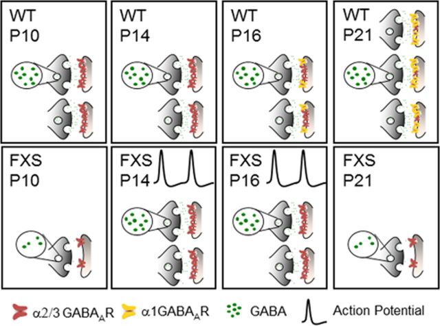
Fragile X syndrome (FXS) is a debilitating neurodevelopmental disorder thought to arise from disrupted synaptic communication in several key brain regions, including the amygdala, a central processing center for information with emotional and social relevance. Recent studies reveal defects in both excitatory and inhibitory neurotransmission in mature amygdala circuits in Fmr1(-/y) mutants, the animal model of FXS. However, whether these defects are the result of altered synaptic development or simply faulty mature circuits remains unknown. Using a combination of electrophysiological and genetic approaches, we show the development of both presynaptic and postsynaptic components of inhibitory neurotransmission in the FXS amygdala is dynamically altered during critical stages of neural circuit formation. Surprisingly, we observe that there is a homeostatic correction of defective inhibition, which, despite transiently restoring inhibitory synaptic efficacy to levels at or beyond those of control, ultimately fails to be maintained. Using inhibitory interneuron-specific conditional knock-out and rescue mice, we further reveal that fragile X mental retardation protein function in amygdala inhibitory microcircuits can be segregated into distinct presynaptic and postsynaptic components. Collectively, these studies reveal a previously unrecognized complexity of disrupted neuronal development in FXS and therefore have direct implications for establishing novel temporal and region-specific targeted therapies to ameliorate core amygdala-based behavioral symptoms.


Figures

Figure 1.
Figure 1.
The development of inhibitory neurotransmission is dynamically altered in the Fmr1−/y mutant BLA. A, Recordings of sIPSCs recorded from BLA excitatory neurons. B–D, Histograms of pooled data for sIPSC biophysical parameters: amplitude (B), frequency (C), and decay constant (D). Mean ± SEM, sample sizes, and p values (Student's two-tailed t test vs control) are given in Table 1. *p < 0.05; **p < 0.01. E–G, Histograms of pooled data for mIPSC biophysical parameters: amplitude (E), frequency (F), and decay constant (G). Mean ± SEM, sample sizes, and p values are given in Table 1. Note that dynamic fluctuations in inhibitory function are observed over development.
Figure 2.
Figure 2.
Dynamic changes in the levels of synaptic GABA concentration at inhibitory synapses in the Fmr1−/y BLA. The ability of TPMPA to attenuate the amplitude of mIPSCs is inversely proportional to the concentration of GABA localized to the synapse. A, Averages of >150 mIPSC events recorded in the absence (black sweeps for control; gray sweeps for Fmr1−/y) or presence (red traces) of 100 mm TPMPA. B, Histogram of pooled data of changes in mIPSC amplitude in the presence of TPMPA. Note that TPMPA has a significantly greater efficacy at P10 (*p < 0.05) and P21 (**p < 0.01) but not at P14 or P16 in Fmr1−/y. Mean ± SEM change in mIPSC amplitude, sample sizes, and p values are given in Table 2.
Figure 3.
Figure 3.
Developmental increase of GABA transporter function and expression. A, B, Averages of >150 sIPSC events normalized for amplitude before (black and gray traces for control and Fmr1−/y, respectively) and after (red traces) the application of 1 μm NO-711. A, At P10, no significant differences in decay constant were observed in either genotype in the presence of NO-711. B, NO-711 significantly increased the decay constants of both control and Fmr1−/y mutant sIPSCs at P21. C, NO-711 enhancement as a measure of the percentage difference in decay constant before and after treatment with NO-711. Note that NO-711 significantly increased the decay constant of P21 Fmr1−/y sIPSCs to a greater degree than in control sIPSCs (*p < 0.05). All decay constant and NO-711 enhancement values ± SEM, sample sizes, and p values are listed in Table 2. Immunohistochemical detection of GAT1 expression in control (D) and Fmr1−/y KOs (E). LA, Lateral amygdala; PirC, piriform cortex. Scale bars, 250 μm.
Figure 4.
Figure 4.
GABAAR subunit-selective pharmacology of sIPSCs is disrupted in the Fmr1−/y mutant BLA. A, Sensitivity to the α1 GABAAR agonist zolpidem is reduced in Fmr1−/y mutants at postnatal ages. sIPSCs were recorded in the presence of 200 nm zolpidem, an α1 GABAAR agonist that acts to lengthen the decay constant. “Zolpidem Enhancement” measures the percentage difference between the decay constant in the presence of zolpidem and that measured in the absence of the drug. Control responses to zolpidem increased steadily throughout development, consistent with the developmentally regulated increase in α1 GABAARs (black line). In contrast, sIPSCs from Fmr1−/y mutant principal neurons showed no increase in zolpidem sensitivity (gray line). *p < 0.05; **p < 0.01. B, The developmental profile of clonazapam-induced pharmacological changes is also disrupted. Overall, clonazepam sensitivity decreases throughout development in control, consistent with the developmental decrease in α3-containing GABAARs. Note that this trend is disrupted in Fmr1−/y. *p < 0.05; **p < 0.01. All values ± SEM, sample sizes, and p values are given in Table 2.
Figure 5.
Figure 5.
mIPSC quantal analysis reveals smaller postsynaptic GABAAR patch sizes in Fmr1−/y inhibitory synapses. A, B, Representative mIPSC amplitude histograms for control (A) and Fmr1−/y (B) at P10, P14, P16, and P21. Dashed lines demarcate the least-squares best-fit function. Underlying Gaussian functions are shown in colored lines. C, Summary of the number of deconvolved Gaussians across cells for control and Fmr1−/y at each corresponding age. Note that the average number of Gaussians was only significantly different between genotypes at P10 (*p < 0.05) and P21 (**p < 0.01).
Figure 6.
Figure 6.
Peak-scaled nonstationary noise analysis suggests significantly higher unit currents and lower numbers of GABAARs in Fmr1−/y at P10 and P21 but not at P14 or P16. A, B, Current variance (σ2) and mean current (Im) plots for control (A) and Fmr1−/y (B) mIPSCs at P21. Solid lines indicate the best fit of the parabolic function σ2 = iImIm2/N. C, D, Mean estimated values for unit current (i; C) and the number of GABAARs (N; D) over development. **p < 0.01, Kolmorogov–Smirnoff test.
Figure 7.
Figure 7.
Comparison of biophysical parameters for sIPSCs in conditional KO and rescue animals to control and full KO animals. Histograms of pooled data of sIPSCs recorded from P10 (A), P14 (B), and P21 (C) time points. Amplitude, frequency, and decay constant of sIPSCs recorded from control (black bars), full KO (Fmr1−/y; gray bars), control animals with cre driver (Dlx5/6Cre; purple bars), conditional KOs (Dlx5/6Cre;Fmr1cKO; red bars), and conditional rescues (Dlx5/6Cre;Fmr1cON; green bars) cells. Sensitivity to zolpidem assessment as a measure of percentage enhancement indicates that Dlx5/6Cre;Fmr1cKO BLA principal neurons show no preference at all ages. *p < 0.05; **p < 0.001; ***p < 0.001, significantly different from control.
Figure 8.
Figure 8.
Summary of altered developmental inhibitory synaptic processes in FXS. The top row depicts development across the four postnatal time points in the wild-type BLA, and the bottom row shows the case of FXS. Our results suggest that at least five processes are dynamically disrupted in the development of inhibitory neurotransmission in the FXS BLA. First, changes in IPSC frequency suggest that there may be alterations in the number of synapses throughout development, with more synapses becoming active at P14 and P16. Second, because the frequency of inhibitory synaptic inputs is elevated beyond control levels at P14–P16 only when action potentials are not blocked, BLA interneurons in Fmr1−/y may have higher firing rates. Third, presynaptic mechanisms governing the amount of GABA in the synapse are transiently upregulated, restoring the amount of synaptic GABA to control levels at P14–P16. Fourth, there is a severe defect in the process of GABAAR maturation in the FXS BLA, in which mature α1 GABAARs fail to become functional, whereas immature α3 GABAARs are retained at mature ages. Finally, the number of GABAARs increases to control levels at P14–P16. However, these compensatory changes fail to be maintained, and inhibitory defects are again present in the mature FXS BLA.

Similar articles

Cited by

References

    1. Avila MA, Real MA, Guirado S. Patterns of GABA and GABA transporter-1 immunoreactivities in the developing and adult mouse brain amygdala. Brain Res. 2011;1388:1–11. doi: 10.1016/j.brainres.2011.02.093. - DOI - PubMed
    1. Barberis A, Lu C, Vicini S, Mozrzymas JW. Developmental changes of GABA synaptic transient in cerebellar granule cells. Mol Pharmacol. 2005;67:1221–1228. doi: 10.1124/mol.104.006437. - DOI - PubMed
    1. Bear MF, Huber KM, Warren ST. The mGluR theory of fragile X mental retardation. Trends Neurosci. 2004;27:370–377. doi: 10.1016/j.tins.2004.04.009. - DOI - PubMed
    1. Centonze D, Rossi S, Mercaldo V, Napoli I, Ciotti MT, De Chiara V, Musella A, Prosperetti C, Calabresi P, Bernardi G, Bagni C. Abnormal striatal GABA transmission in the mouse model for the fragile X syndrome. Biol Psychiatry. 2008;63:963–973. doi: 10.1016/j.biopsych.2007.09.008. - DOI - PubMed
    1. Comery TA, Harris JB, Willems PJ, Oostra BA, Irwin SA, Weiler IJ, Greenough WT. Abnormal dendritic spines in fragile X knockout mice: maturation and pruning deficits. Proc Natl Acad Sci U S A. 1997;94:5401–5404. doi: 10.1073/pnas.94.10.5401. - DOI - PMC - PubMed

Publication types