. 2013 Aug;27(8):3017-29.
doi: 10.1096/fj.12-224824. Epub 2013 Apr 24.

Monocytes mediate metastatic breast tumor cell adhesion to endothelium under flow

Affiliations

Monocytes mediate metastatic breast tumor cell adhesion to endothelium under flow

Shankar J Evani et al. FASEB J. 2013 Aug.

Abstract

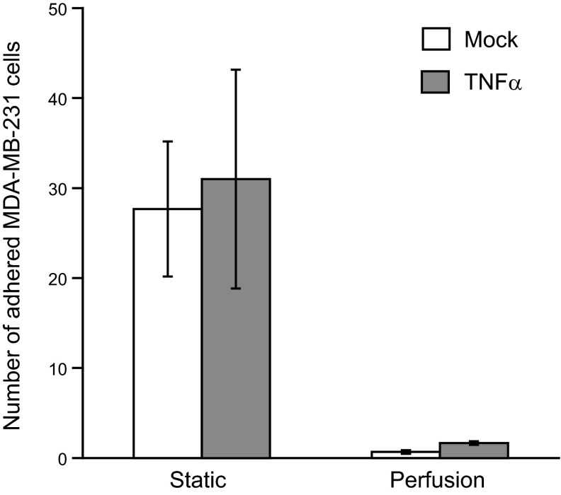
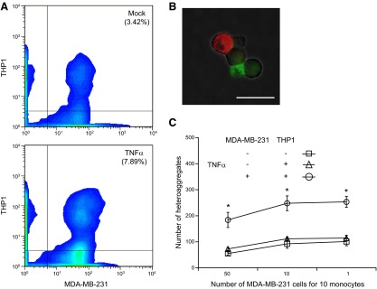
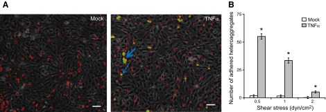
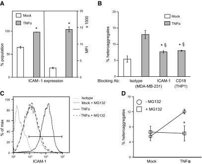
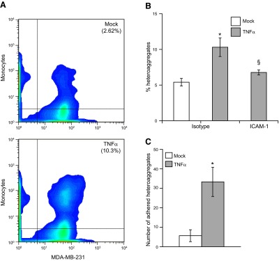
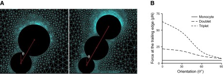
Endothelial adhesion is necessary for the hematogenous dissemination of tumor cells. However, the metastatic breast tumor cell MDA-MB-231 does not bind to the endothelium under physiological flow conditions, suggesting alternate mechanisms of adhesion. Since monocytes are highly represented in the tumor microenvironment, and also bind to endothelium during inflammation, we hypothesized that the monocytes assist in the arrest of MDA-MB-231 on the endothelium. Using in vitro models of the dynamic shear environment of the vasculature, we show that TNF-α-activated THP1/primary human monocytes and MDA-MB-231 cells form stable aggregates, and that the monocytes in these aggregates mediate the adhesion of otherwise nonadherent MDA-MB-231 cells to inflamed endothelium under flow (55±2.4 vs. 1.7±0.82 at a shear stress of 0.5 dyn/cm(2), P<0.01). We also show that the hydrodynamic forces determine the size and orientation of aggregates adhered to the endothelium, and strongly favor the attachment of small aggregates with tumor cells downstream of flow (74-86% doublets at 0.5-2 dyn/cm(2), P<0.01). The 5-fold up-regulation of ICAM-1 on TNF-α-activated MDA-MB-231 cells through the Nf-κB pathway was found to be critical in MDA-MB-231-monocyte aggregation and endothelial adhesion. Our results demonstrate that, under inflammatory conditions, monocytes may serve to disseminate tumor cells through circulation, and the tumor-monocyte-endothelial axis may represent a new therapeutic target to reduce cancer metastasis.

Keywords: MDA-MB-231; aggregation; shear.


Figures

Figure 1.
Figure 1.
Adhesion of MDA-MB-231 cells to endothelium. Untreated (mock) or TNF-α-treated (10 ng/ml for 6 h, TNF-α) MDA-MB-231 cells at a concentration of 106 cells/ml were perfused at a constant flow rate, corresponding to a wall shear stress of 0.5 dyn/cm2 for 2 min over a confluent monolayer of HAECs activated with TNF-α (10 ng/ml for 6 h). The flow was stopped, and the cells were allowed to adhere onto the HAECs for 4 min (static group). Nonspecifically adhered cells were washed off by perfusing HBSS buffer at 2 dyn/cm2 for 5 min. In other experiments, the flow was not stopped, and the cells were perfused continuously (perfusion group). The firmly adhered cells were counted from five different fields of view for each experiment. Results are expressed as means ± sd of a representative experiment performed in triplicate; experiments were performed 3 times.
Figure 2.
Figure 2.
Aggregation of MDA-MB-231 and THP1 cells. Prestained THP1 cells (PKH-26, FITC channel) and MDA-MB-231 cells (PKH-67, PE channel) were either untreated (mock) or treated with 10 ng/ml TNF-α (TNF-α) for 6 h, and resuspended to 2 × 10⁶ cells/ml in corresponding complete medium with 2% BSA. Equal volumes of the cell suspensions were incubated for 20 min with rocking at 37°C and gently mixed with pipette to disaggregate any nonspecific aggregates. Suspensions (80 μl) were subjected to a shear stress of 5 dyn/cm2 for 2 min in a cone-and-plate viscometer at 37°C. Samples (20 μl) were immediately fixed and analyzed by flow cytometry. A) MDA-MB-231/THP1 heteroaggregates were quantified as the number of events that were positive for fluorescence in both FITC and PE channels. B) Fluorescence microscopic images of aggregates in mock- and TNF-α-treated samples. Scale bar = 20 μm. C) MDA-MB-231/THP1 heteroaggregates in suspension containing different initial ratios of MDA-MB-231:THP1 cells. Results are expressed as means ± sd of a representative experiment performed in triplicate; experiments were performed 3 times. *P < 0.01 vs. untreated controls.
Figure 3.
Figure 3.
Adhesion of MDA-MB-231/THP1 heteroaggregates to endothelium. A suspension of MDA-MB-231/THP1 aggregates was prepared as described in Fig. 2. The suspension was perfused at 0.5, 1 and 2 dyn/cm2 on top of TNF-α-activated HAECs, and the number of adherent MDA-MB-231 cells was surveyed by fluorescence and bright-field microscopy. A) In cell suspension not treated with TNF-α, only THP1 cells (red) bind to the endothelium, but in cell suspension treated with TNF-α, MDA-MB-231/THP1 heteroaggregates (green-red) bind to the endothelium. Arrows represent aggregates attached to endothelium. Scale bars = 20 μm. B) Number of adherent MDA-MB-231 cells (in heteroaggregates) was calculated from 5 fields of view. Results are expressed as means ± sd of a representative experiment performed in triplicate; experiments were performed 3 times. *P < 0.01 vs. untreated controls.
Figure 4.
Figure 4.
Size and orientation of MDA-MB-231/THP1 heteroaggregates attached to the endothelium. Untreated or TNF-α-treated MDA-MB-231/THP1 heteroaggregates were perfused over activated HAECs as described in Fig. 3. A) Numbers of doublets, containing 1 MDA-MB-231 cell and 1 THP1 cell, or triplets and larger aggregates that attached stably to the endothelium were counted. B) Representative instantaneous velocity profiles of THP1 cells, MDA-MB-231/THP1 doublets, and triplet heteroaggregates of 2 MDA-MB-231 cells and a THP1 cell rolling on the endothelium at a wall shear rate of 1 dyn/cm2. C) Orientation of MDA-MB-231 cells with respect to the flow direction in a stably attached aggregate at a wall shear rate of 1 dyn/cm2. Fluorescence microscopic images of THP1 (red) and MDA-MB-231 (green) were merged, with phase-contrast image of endothelial cells in the background. Scale bar = 20 μm. Center red circle represents a THP1 cell; inner green circles and outer blue circles represent the locations of MDA MB-231 cells during initial tethering and final stable attachment, respectively. Angle between direction of flow and the location of MDA-MB-231 cells with respect to THP1 in heteroaggregates was calculated from 5 fields of view. Results are expressed as means ± sd of a representative experiment performed in triplicate; experiments were performed 3 times.
Figure 5.
Figure 5.
Mechanism of MDA-MB-231/THP1 aggregation. A) Untreated or TNF-α-treated MDA-MB-231 cells were fixed, stained with PE-conjugated ICAM-1 antibody or the corresponding isotype control, and analyzed by flow cytometry. Results are averages of a representative experiment run in triplicate; experiment was repeated 2 times. *P < 0.01 vs. mock treatment. B) TNF-α-activated MDA-MB-231 and THP1 cells were blocked with anti-ICAM-1 or anti-CD18 antibody, respectively, or with the corresponding isotype controls, before preparing and quantifying heteroaggregates as described in Fig. 2. *P < 0.01 vs. TNF-α-treated cells without blocking; §P < 0.01 vs. mock-treated control. C, D) MDA-MB-231 cells were preincubated with 0 or 20 μM of MG132 for 30 min before TNF-α treatment. ICAM-1 expression levels were analyzed by flow cytometry (C), and the aggregation experiments (D) were performed as described above. Results are expressed as means ± sd of a representative experiment performed in triplicate; experiments were performed 3 times. *P < 0.01 vs. no MG132.
Figure 6.
Figure 6.
Mechanism of MDA-MB-231/THP1 adhesion to endothelium. A) MDA-MB-231 cells were blocked with anti-ICAM-1 antibody, and a suspension of MDA-MB-231/THP1 aggregates was prepared as described in Fig. 2. Aggregates were perfused on top of endothelium at 1 dyn/cm2, and the number of aggregates attaching stably to the endothelium was quantified as described in Fig. 3. B) MDA-MB-231 cells were preincubated with 0 or 20 μM of MG132 for 30 min before TNF-α treatment. A suspension of MG132-treated MDA-MB-231 and THP1 aggregates was perfused on top of endothelium, and stable aggregates attached to the endothelium were quantified. *P < 0.01 vs. untreated controls.
Figure 7.
Figure 7.
MDA-MB-231/primary monocyte heteroaggregation and endothelial adhesion. A) Aggregation experiments and quantification were identical to those described in Fig. 2, except that human monocytes were used. Formaldehyde-fixed MDA-MB-231/primary monocyte heteroaggregates were quantified as the percentage of events that were positive for fluorescence in both FITC and PE channels. B) TNF-α-activated MDA-MB-231 cells were blocked with anti-ICAM-1 or the corresponding isotype controls, before heteroaggregates were prepared with human monocytes and quantified by flow cytometry. *P < 0.05 vs. untreated controls. C) Suspension containing MDA-MB-231/monocyte aggregates was perfused at 1 dyn/cm2 on top of TNF-α-activated HAECs, and the number of adherent MDA-MB-231 cells was surveyed by fluorescence microscopy in 5 fields of view. Results are means ± sd of a representative experiment performed in triplicate; experiments were repeated 3 times. *P < 0.01 vs. mock treatment.
Figure 8.
Figure 8.
Estimation of detachment force at the monocyte-endothelial interface of heteroaggregates due to fluid flow. A) Geometric configurations of MDA-MB-231/monocyte heteroaggregates attached to endothelial surface under laminar shear flow. Configurations include a MDA-MB-231/THP1 doublet and a triplet consisting of 2 MDA-MB-231 cells and a monocyte. Angle θ represents the orientation of the MDA-MB-231 cell that is attached to the monocyte with respect to the vertical axis. B) Reaction force experienced at the trailing edge of the monocyte–endothelial contact area.

Similar articles

Cited by

References

    1. Valastyan S., Weinberg R. A. (2011) Tumor metastasis: molecular insights and evolving paradigms. Cell 147, 275–292 - PMC - PubMed
    1. Liang S., Slattery M. J., Wagner D., Simon S. I., Dong C. (2008) Hydrodynamic shear rate regulates melanoma-leukocyte aggregation, melanoma adhesion to the endothelium, and subsequent extravasation. Ann. Biomed. Eng. 36, 661–671 - PMC - PubMed
    1. Konstantopoulos K., Thomas S. N. (2009) Cancer cells in transit: the vascular interactions of tumor cells. Annu. Rev. Biomed. Eng. 11, 177–202 - PubMed
    1. Jadhav S., Bochner B., Konstantopoulos K. (2001) Hydrodynamic shear regulates the kinetics and receptor specificity of polymorphonuclear leukocyte-colon carcinoma cell adhesive interactions. J. Immunol. 167, 5986–5993 - PubMed
    1. Geng Y., Narasipura S., Seigel G. M., King M. R. (2010) Vascular recruitment of human retinoblastoma cells by multi-cellular adhesive interactions with circulating leukocytes. Cell. Mol. Bioeng. 3, 361–368 - PMC - PubMed

Publication types

MeSH terms

Substances