Review
. 2013 Jun;13(3):153-64.
doi: 10.1136/practneurol-2013-000557. Epub 2013 Apr 24.

Mimics and chameleons in motor neurone disease

Affiliations
Free PMC article
Review

Mimics and chameleons in motor neurone disease

Martin R Turner et al. Pract Neurol. 2013 Jun.
Free PMC article

Abstract

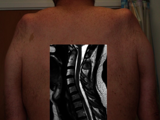
The progression of motor neurone disease (MND) is currently irreversible, and the grave implications of diagnosis naturally fuels concern among neurologists over missing a potential mimic disorder. There is no diagnostic test for MND but in reality there are few plausible mimics in routine clinical practice. In the presence of a progressive pure motor disorder, signs such as florid fasciculations, bilateral tongue wasting, the 'split hand', head drop, emotionality, and cognitive or behavioural impairment carry high positive predictive value. MND is clinically heterogeneous, however, with some important chameleon-like presentations and considerable variation in clinical course. Lack of confidence about the scope of such variation, or an approach to diagnosis emphasising investigations over clinical common sense, has the potential to exacerbate diagnostic delay in MND and impede timely planning of the care which is essential to maximising quality of life.

Keywords: MOTOR NEURON DISEASE.


Figures

Figure 1
Figure 1
The diagnostic pathway in motor neurone disease (MND). There is an average delay of 1 year between symptom onset and diagnosis. There is a powerful relationship between rate of progression and speed of diagnosis. ALS, amyotrophic lateral sclerosis; GP, general practitioner; UMN, upper motor neurone.
Figure 2
Figure 2
Multifocal motor neuropathy with conduction block. Hyperintense swollen right-sided nerve roots on cervical spine coronal MRI (left panel, modified with permission from71). It typically causes asymmetrical distal weakness, often involving finger extensors initially, but can progress to wasting and fixed flexion deformity, as in this patient, several years after symptom onset (right panel; for other images see72).
Figure 3
Figure 3
Marked bilateral tongue and hand wasting in Kennedy's disease. The clue to this case not being motor neurone disease was the very slow progression, lack of upper motor neurone signs, gynaecomastia and chin fasciculations.
Figure 4
Figure 4
Typical bilateral medial forearm and quadriceps (right leg only in this case) wasting of inclusion body myositis. There is a characteristic rimmed vacuole within a muscle fibre histopathologically (with acknowledgement to Dr Waney Squier).
Figure 5
Figure 5
Flexion cervical spine MRI in Hirayama disease showing expansion of the dural venous plexus with presumed chronic ischaemic damage preferentially involving the anterior horn cells supplying the distal arm.
Figure 6
Figure 6
This patient (shown from behind) presented with slowly progressive weakness and wasting of the shoulder girdles, neurophysiological evidence of denervation and slightly raised serum creatine kinase, all compatible with the ‘flail arm’ variant of motor neurone disease. MRI (inset) of the cervical spine showed expansion of the dural space posteriorly with forward compression of the spinal cord and presumed chronic damage to the anterior horns cells. His progression arrested spontaneously.
Figure 7
Figure 7
Radiation-induced lumbar radiculopathy. This patient presented with a slowly progressive right leg weakness and wasting more than 30 years after radiotherapy to the right para-aortic nodes for testicular cancer (radiotherapy tattoos shown circled over abdomen).

Similar articles

Cited by

References

    1. Davies Z, Turner MR. Public awareness of motor neuron disease. Amyotroph Lateral Scler 2010;11:490–1 - PMC - PubMed
    1. Matthews WB. Practical neurology. 2nd edn Philadelphia: F.A. Davis Company, 1970
    1. Turner MR, Bowser R, Bruijn L, et al. Mechanisms, models and biomarkers in amyotrophic lateral sclerosis. ALS and Frontotemporal Degeneration. 2013;14 (suppl 1): 19–32 - PMC - PubMed
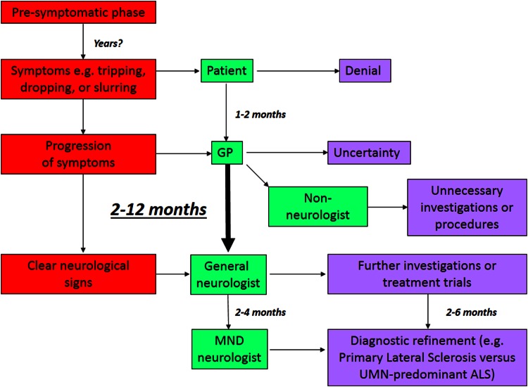
    1. Mitchell JD, Callagher P, Gardham J, et al. Timelines in the diagnostic evaluation of people with suspected amyotrophic lateral sclerosis (ALS)/motor neuron disease (MND)—a 20-year review: Can we do better? Amyotroph Lateral Scler 2010;11:537–41 - PubMed
    1. Kiernan MC, Vucic S, Cheah BC, et al. Amyotrophic lateral sclerosis. Lancet 2011;377:942–55 - PubMed

Publication types