. 2013 May 21;127(20):2038-46.
doi: 10.1161/CIRCULATIONAHA.112.000116. Epub 2013 Apr 24.

Monocyte-directed RNAi targeting CCR2 improves infarct healing in atherosclerosis-prone mice

Affiliations

Monocyte-directed RNAi targeting CCR2 improves infarct healing in atherosclerosis-prone mice

Maulik D Majmudar et al. Circulation. .

Abstract

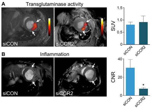
Background: Exaggerated and prolonged inflammation after myocardial infarction (MI) accelerates left ventricular remodeling. Inflammatory pathways may present a therapeutic target to prevent post-MI heart failure. However, the appropriate magnitude and timing of interventions are largely unknown, in part because noninvasive monitoring tools are lacking. Here, we used nanoparticle-facilitated silencing of CCR2, the chemokine receptor that governs inflammatory Ly-6C(high) monocyte subset traffic, to reduce infarct inflammation in apolipoprotein E-deficient (apoE(-/-)) mice after MI. We used dual-target positron emission tomography/magnetic resonance imaging of transglutaminase factor XIII (FXIII) and myeloperoxidase (MPO) activity to monitor how monocyte subset-targeted RNAi altered infarct inflammation and healing.

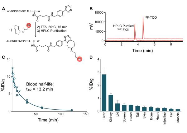
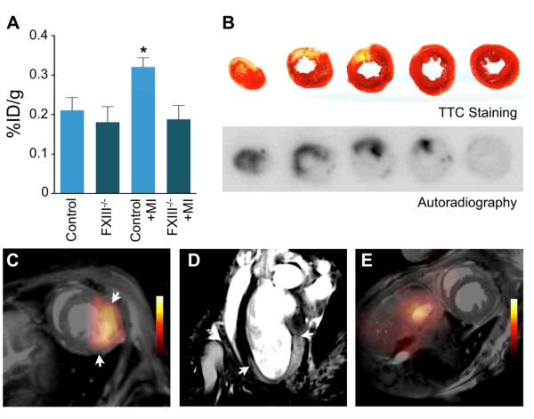
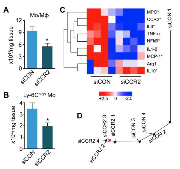
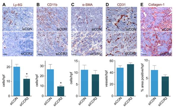
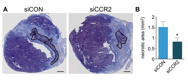
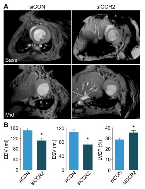
Methods and results: Flow cytometry, gene expression analysis, and histology revealed reduced monocyte numbers and enhanced resolution of inflammation in infarcted hearts of apoE(-/-) mice that were treated with nanoparticle-encapsulated siRNA. To follow extracellular matrix cross-linking noninvasively, we developed a fluorine-18-labeled positron emission tomography agent ((18)F-FXIII). Recruitment of MPO-rich inflammatory leukocytes was imaged with a molecular magnetic resonance imaging sensor of MPO activity (MPO-Gd). Positron emission tomography/magnetic resonance imaging detected anti-inflammatory effects of intravenous nanoparticle-facilitated siRNA therapy (75% decrease of MPO-Gd signal; P<0.05), whereas (18)F-FXIII positron emission tomography reflected unimpeded matrix cross-linking in the infarct. Silencing of CCR2 during the first week after MI improved ejection fraction on day 21 after MI from 29% to 35% (P<0.05).

Conclusion: CCR2-targeted RNAi reduced recruitment of Ly-6C(high) monocytes, attenuated infarct inflammation, and curbed post-MI left ventricular remodeling.

Keywords: RNA, small interfering; heart failure; inflammation; magnetic resonance imaging; molecular imaging; monocytes; myocardial infarction; positron-emission tomography; ventricular remodeling.


Figures

Figure 1
Figure 1. 18F-FXIII PET agent
A, Schematic representation of agent synthesis. Factor XIII-tetrazine (FXIII-Tz) was reacted with prosthetic group 18F-trans-cyclooctene (18F-TCO) to give 18F-FXIII. B, Analytical HPLC chromatographs 18F-TCO and 18F-FXIII following preparative scale HPLC purification. C, Blood half-life of 18F-FXIII (n=5). D, Biodistribution of 18F-FXIII PET agent 75 minutes after intravenous administration (n=5). Data are presented as mean±SEM.
Figure 2
Figure 2. 18F-FXIII/MPO-Gd PET/MRI in MI
A, Scintillation counting in hearts of control B6 mice with and without MI compared to FXIII−/− mice (day 4 after MI, n=4-5 per group). Data are mean±SEM, *p<0.05; p-value was tested between B6 mice with MI and control B6 mice. B, TTC staining of infarcted tissue (yellow) and viable myocardium (red) correlated with 18F-FXIII signal on autoradiography in B6 mouse on day 4 after MI. C-D, In vivo dual target molecular PET/MRI of wound healing and myocardial inflammation in a wild type B6 mouse on day 4 after coronary ligation. The short axis (C) shows 18F-FXIII PET signal in MI fused to MR image. A representative long axis MR image (D) shows signal enhancement in the infarct area 90 minutes after intravenous MPO-Gd injection. Arrows indicate infarct borders. E, PET/MRI of control mouse without coronary ligation.
Figure 3
Figure 3. Monocytes are not a major source of FXIII in MI
A, Scintillation counting of infarcted hearts from mice with or without clodronate liposome (Clo-Lip) depletion of monocytes and macrophages after injection of 18F-FXIII (n=4-5 per group). B, Comparison of 18F-FXIII signal on autoradiography between control mouse (MI) and mouse treated with Clo-Lip (MI + Clo-Lip). Data are mean±SEM, *p<0.05. %ID/g: percent injected dose per gram tissue.
Figure 4
Figure 4. Treatment with siCCR2 reduces inflammatory monocytes in the heart
A, Flow cytometric analysis of 4 day old infarcts shows decreased CD11b+ lineage myeloid cells (Mo/Mϕ). Dot plots and gating strategy are shown in supplementary Figure 1. B, Number of Ly-6Chigh monocytes in infarcts. Data are mean±SEM, n=4 per group, *p<0.05. C, Heat map of genes in infarcted myocardium (n=4 per group). Each row of the heat map represents a gene while each column represents an experimental treatment group (labeled at the bottom). The color scale represents the level of gene expression, with red indicating an increase in gene expression and blue indicating a decrease in gene expression. Data underwent z-score transformation for display. D, Distance tree showing the relative location and clustering of treatment cohorts.
Figure 5
Figure 5. siCCR2 treatment effects on wound healing
A-E, Immunohistochemical evaluation of the infarct after siCCR2 or siCON treatment for neutrophils (A, Ly-6G), myeloid cells (B, CD11b), myofibroblasts (C, α-SMA), neovessels (D, CD31), and collagen deposition (E, collagen-1). Images depict border zone of the infarct. n=4 mice per group and n=4 high power fields (hpf) /mouse. Data are mean±SEM, *p<0.05. Scale bar indicates 25μm.
Figure 6
Figure 6. CCR2 silencing reduces residual necrotic debris
A, Masson trichrome stain with residual necrotic debris in infarct outlined in black. B, Comparison of residual necrotic debris between siCON and siCCR2 treated animals (4 mice per group). Data are mean±SEM, *p<0.05. Scale bar indicates 0.5 mm.
Figure 7
Figure 7. Dual target molecular PET/MRI after therapeutic RNA silencing of CCR2
A, Short axis PET/MRI images after respective siRNA treatment. Bar graph of standard uptake value (SUV) shows no difference in 18F-FXIII PET signal between treatment groups (n=5 per group). B, Short axis MRI showing MPO-Gd signal enhancement in siCON and reduced enhancement in siCCR2 treated mice. Arrows indicate infarct area. The bar graph shows contrast-to-noise ratio (CNR). Data are mean±SEM, *p<0.05.
Figure 8
Figure 8. CCR2 silencing reduces adverse ventricular remodeling
A, Basal and mid-ventricular short axis cine MR images on day 21 after MI. B, Bar graphs depict differences in left ventricular remodeling between siCON and siCCR2 treated cohorts (n=5 per group). Data are mean±SEM, *p<0.05. EDV: end-diastolic volume; ESV: end-systolic volume; LVEF: left ventricular ejection fraction.

Comment in

  • Blinding the monocytes to protect the heart.
    Charo IF. Charo IF. Circulation. 2013 May 21;127(20):2006-8. doi: 10.1161/CIRCULATIONAHA.113.003045. Epub 2013 Apr 24. Circulation. 2013. PMID: 23616628 Free PMC article. No abstract available.

Similar articles

Cited by

References

    1. Frangogiannis NG, Smith CW, Entman ML. The inflammatory response in myocardial infarction. Cardiovasc Res. 2002;53:31–47. - PubMed
    1. Frantz S, Bauersachs J, Ertl G. Post-infarct remodelling: contribution of wound healing and inflammation. Cardiovasc Res. 2009;81:474–481. - PMC - PubMed
    1. Lambert JM, Lopez EF, Lindsey ML. Macrophage roles following myocardial infarction. Int J Cardiol. 2008;130:147–158. - PMC - PubMed
    1. French BA, Kramer CM. Mechanisms of Post-Infarct Left Ventricular Remodeling. Drug Discov Today Dis Mech. 2007;4:185–196. - PMC - PubMed
    1. Lindsey ML, Mann DL, Entman ML, Spinale FG. Extracellular matrix remodeling following myocardial injury. Ann Med. 2003;35:316–326. - PubMed

Publication types

MeSH terms