. 2013 Apr 17:4:48.
doi: 10.3389/fendo.2013.00048. eCollection 2013.

GABA and Glutamate Uptake and Metabolism in Retinal Glial (Müller) Cells

Affiliations

GABA and Glutamate Uptake and Metabolism in Retinal Glial (Müller) Cells

Andreas Bringmann et al. Front Endocrinol (Lausanne). .

Abstract

Müller cells, the principal glial cells of the retina, support the synaptic activity by the uptake and metabolization of extracellular neurotransmitters. Müller cells express uptake and exchange systems for various neurotransmitters including glutamate and γ-aminobutyric acid (GABA). Müller cells remove the bulk of extracellular glutamate in the inner retina and contribute to the glutamate clearance around photoreceptor terminals. By the uptake of glutamate, Müller cells are involved in the shaping and termination of the synaptic activity, particularly in the inner retina. Reactive Müller cells are neuroprotective, e.g., by the clearance of excess extracellular glutamate, but may also contribute to neuronal degeneration by a malfunctioning or even reversal of glial glutamate transporters, or by a downregulation of the key enzyme, glutamine synthetase. This review summarizes the present knowledge about the role of Müller cells in the clearance and metabolization of extracellular glutamate and GABA. Some major pathways of GABA and glutamate metabolism in Müller cells are described; these pathways are involved in the glutamate-glutamine cycle of the retina, in the defense against oxidative stress via the production of glutathione, and in the production of substrates for the neuronal energy metabolism.

Keywords: GABA; Müller cells; glutamate; recycling; retina; retinal pathology.


Figures

Figure 1
Figure 1
Müller cells span the entire thickness of the neuroretina. Schematic drawing of the cellular constituents and basic neuronal circuits of a human retina. The inner retina contact the vitreal cavity (top) and contains the following retinal layers: nerve fiber layer (NFL), ganglion cell layer (GCL), inner plexiform layers (IPL), and inner nuclear layer (INL). The outer retina is directed to the outer surface of the eye and contains the following retinal layers: outer plexiform layer (OPL), outer nuclear layer (ONL), the subretinal space containing the photoreceptor segments (PRS), and the retinal pigment epithelium (RPE). The outer retina is supplied with oxygen and nutrients by the blood vessels (BV) in the choroid (Chor), while the inner retina is supplied by intraretinal vessels. Astrocytes (as) are localized in the NFL/GCL. The perikarya of Müller cells are localized in the INL. From the perikaryon, two stem processes of Müller cells extend toward both surfaces of the neuroretina. The funnel-shaped endfeet of Müller cells form (in association with a basement membrane) the inner surface of the retina. In the OPL and IPL, side branches which form perisynaptic membrane sheaths originate at the stem processes. In the ONL, the stem process of Müller cells forms membraneous sheaths which envelop the perikarya of rods (r) and cones (c). Microvilli of Müller cells extend into the subretinal space. ac, amacrine cell; bc, bipolar cell; rgc, retinal ganglion cell.
Figure 2
Figure 2
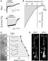
Electrogenic GABA transport in Müller cells. The GABA-evoked membrane currents were recorded in freshly isolated Müller cells from man (A) and guinea pigs (A–C). (A) Extracellular administration of GABA (100 μM) evokes two kinds of inward currents in a human Müller cell: a transient, rapidly inactivating chloride current mediated by GABAA receptors (arrow) and a sustained current mediated by electrogenic GABA transporters (arrowhead). In guinea pig Müller cells (which lack GABAA receptors), extracellular GABA (1 mM) evokes only a transporter-mediated inward current. The holding potential was −80 mV. (B) Current-voltage relation of the GABA transporter currents in guinea pig Müller cells. The amplitude of the transporter currents is zero near the equilibrium potential of sodium ions (ENa) (when recorded with symmetrical chloride concentration at both sides of the membrane). GABA was administered at a concentration of 100 μM. (C) Subcellular distribution of the GABA transporter currents. The distribution of the currents was determined by focal ejections of GABA (1 mM) onto the following membrane domains of Müller cells: endfoot, inner stem process, soma, inner and outer parts of the outer stem process. (D) Distribution of GAT-1 and GAT-3 immunoreactivities in isolated Müller cells of the guinea pig. Filled arrows, outer stem process. Filled arrowheads, cell soma. Unfilled arrowheads, cell endfoot. Bars, 10 μm. With permission from Biedermann et al. (2002, 2004).
Figure 3
Figure 3
Recycling of amino acid neurotransmitters in the outer plexiform (synaptic) layer of the mammalian retina. The ribbon synapse of a photoreceptor cell (PC) synthesizes glutamate which is continuously released during darkness. The postsynaptic elements are dendrites of bipolar (BC) and horizontal cells (HC). Horizontal cells release GABA which is formed from glutamate. The synaptic complexes are surrounded by Müller cell sheets; the right side shows neurotransmitter uptake systems and some metabolization ways of Müller cells. Glutamate, GABA, and ammonia (NH4+) are transported into the Müller cell and transformed to glutamine and α-ketoglutarate (α-KG). Glutamine is released from Müller cells and serves as precursor for the transmitter synthesis in neurons (glutamate-glutamine cycle). Lactate, alanine, pyruvate, α-ketoglutarate, and glutamine are utilized by neurons as substrates for their energy metabolism. The mitochondrial enzyme GABA transaminase (GABA-T) catalyzes the formation of glutamate from 2-oxoglutarate (2OG), coupled to a conversion of GABA to succinate semialdehyde (SSA). Another metabolic way is the production of reduced glutathione (GSH) which is an intracellular antioxidant, released from Müller cells and taken up by neurons under oxidative stress conditions. AcCoA, acetyl coenzyme A; ALAT, alanine aminotransferase; CGA, cystine-glutamate antiporter; CyT, cystine transporter; EAAC1, excitatory amino acid carrier 1; EAAT5, excitatory amino acid transporter 5; GAD, glutamic acid decarboxylase; GAT, GABA transporter; GLAST, glutamate-aspartate transporter; GLT-1, glutamate transporter-1; GP, glutathione peroxidase; GR, glutathione reductase; GS, glutamine synthetase; GSSG, glutathione disulfide; LDH, lactate dehydrogenase; PAG, phosphate-activated glutaminase; R, free radicals; TCA, tricarboxylic acid cycle.
Figure 4
Figure 4
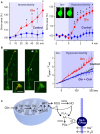
Electrogenic glutamate transport in Müller cells. Whole-cell records of membrane currents were made in freshly isolated cells. (A) Administration of glutamate (1 mM) to a Müller cell of a wild-type mouse evoked inward currents at −80 mV, and outward currents at +50 mV. The inward currents were mediated by the sodium-dependent glutamate uptake, while the outward currents were mediated by the anionic conductance of the glutamate transporter. (The amplitude of the anionic conductance was increased by replacing extracellular chloride with thiocyanate.) In a cell of a GLAST knockout mouse, glutamate did not evoke inward currents, whereas outward currents remained. The absence of inward currents may suggest that at the resting membrane potential of murine Müller cells the electrogenic glutamate uptake is mediated by GLAST. The presence of the chloride conductance may suggest that EAAT5 (which has a large chloride conductance and minimal glutamate transport capability) is also expressed in the cells. (B) Current-voltage relation of the glutamate transporter currents in Müller cells of the guinea pig. (The anionic outward conductance is minimal because extracellular chloride instead of thiocyanate was used used to record the currents.) (C) Activation of ionotropic P2X7 receptors (which mediate cation currents and thus a depolarization of the cells) decreases the electrogenic uptake of glutamate in human Müller cells. The uptake currents evoked by glutamate (Glu; 100 μM) were diminished in the presence of the P2X7 receptor agonist 2′-/3′-O-(4-benzoylbenzoyl)-ATP (BzATP; 10 μM). Inhibition of P2X7 activation by KN-62 (1 μM) suppressed the BzATP-evoked current (and cell depolarization), resulting in glutamate uptake currents similar in amplitude as under control conditions. With permission from The Society for Neuroscience (Pannicke et al., 2000) and Sarthy et al. (2005).
Figure 5
Figure 5
Glutamine (Gln) induces mitochondrial dysfunction and Müller cell swelling. Experiments were carried out in retinal slices (A) and isolated Müller cells (B) of the rat. (A) Glutamine (5 mM) in isoosmotic extracellular solution induced a delayed swelling of Müller cells after 10 min of exposure (left) whereas glutamine (5 mM) in hypoosmotic solution (60% of control osmolarity) induced a rapid (within 1 min) swelling of Müller cells (right). The cross-sectional area of Müller cell somata was recorded and is expressed in percent of the value obtained before superfusion of the slices with the solutions (100%). The images display original records of a Müller cell soma obtained before (left) and during (right) superfusion with the glutamine-containing hypoosmotic solution. Scale bar, 5 μm. Significant difference vs. control: *P < 0.05; **P < 0.01. (B) Glutamine induces a dissipation of the mitochondrial membrane potential in Müller cells. The potential was recorded from the endfeet of freshly isolated cells by using the mitochondria-selective potentiometric dye JC-1. Mitochondrial depolarization is indicated by an increase in the green-to-red fluorescence intensity ratio. Left side: Example of a JC-1-loaded cell which was recorded before (left) and during (right) superfusion of a hypoosmotic solution containing glutamine (5 mM). Insets, cell endfoot at higher magnification. Note the decrease in the red fluorescence. Arrow, cell endfoot. Arrowhead, cell soma. Bars, 10 μm. Right: Time-dependent change in the ratio of the green-to-red fluorescence of the JC-1 dye. The fluorescence was recorded in the absence and presence of glutamine (5 mM), before and during the transition of an isoosmotic to a hypoosmotic extracellular solution. Glutamine-induced a faster dissipation of the mitochondrial membrane potential as compared to control. The inhibitor of the mitochondrial permeability transition, cyclosporin A (CsA; 1 μM), prevented the glutamine-induced dissipation of the mitochondrial potential. (C) Hypothetical scheme of the mechanism of glutamine-induced Müller cell swelling. Cytosolic glutamine is transported into the mitochondria and is hydrolyzed there to glutamate (Glu) and ammonia (NH4+) by the action of the phosphate-activated glutaminase (PAG). High ammonia levels stimulate the mitochondrial production of reactive oxygen species (ROS) and induce mitochondrial permeability transition (MPT) that leads to mitochondrial dysfunction, energy failure, and enhanced free radical production. Mitochondria-derived free radicals activate cytosolic enzymes that generate reactive oxygen and nitrogen species, e.g., nitric oxide synthases (NOS). These radicals stimulate the activity of phospholipases A2 (PLA2), that produce arachidonic acid (AA), lipoxygenases, and cyclooxygenases (COX) which generate prostaglandins (PGs). Arachidonic acid and prostaglandins inhibit the sodium-potassium-ATPase resulting in intracellular sodium overload, water influx, and cellular swelling. The energy failure due to mitochondrial dysfunction may contribute to the inhibition of the sodium-potassium-ATPase. With permission from Karl et al. (2011).

References

    1. Agte S., Junek S., Matthias S., Ulbricht E., Erdmann I., Wurm A., et al. (2011). Müller glial cell-provided cellular light guidance through the vital guinea-pig retina. Biophys. J. 101, 2611–261910.1016/j.bpj.2011.09.062 - DOI - PMC - PubMed
    1. Albrecht J., Norenberg M. D. (2006). Glutamine: a Trojan horse in ammonia neurotoxicity. Hepatology 44, 788–79410.1002/hep.21357 - DOI - PubMed
    1. Anccasi R. M., Ornelas I. M., Cossenza M., Persechini P. M., Ventura A. L. (2013). ATP induces the death of developing avian retinal neurons in culture via activation of P2X7 and glutamate receptors. Purinergic Signal. 9, 15–2910.1007/s11302-012-9324-5 - DOI - PMC - PubMed
    1. Bannai S., Tateishi N. (1986). Role of membrane transport in metabolism and function of glutathione in mammals. J. Membr. Biol. 89, 1–810.1007/BF01870891 - DOI - PubMed
    1. Barbour B., Brew H., Attwell D. (1988). Electrogenic glutamate uptake in glial cells is activated by intracellular potassium. Nature 335, 433–43510.1038/335433a0 - DOI - PubMed

LinkOut - more resources