

Automating tumor classification with pixel-by-pixel contrast-enhanced ultrasound perfusion kinetics
- PMID: 23616934
- PMCID: PMC3463888
- DOI: 10.1116/1.3692962
Automating tumor classification with pixel-by-pixel contrast-enhanced ultrasound perfusion kinetics
Abstract
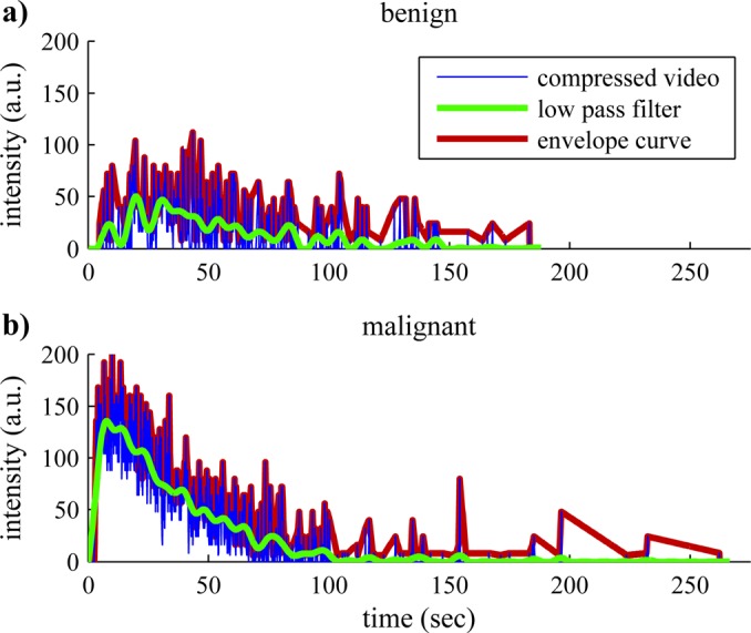
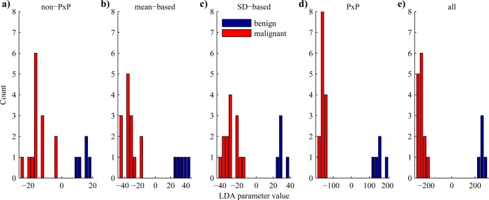
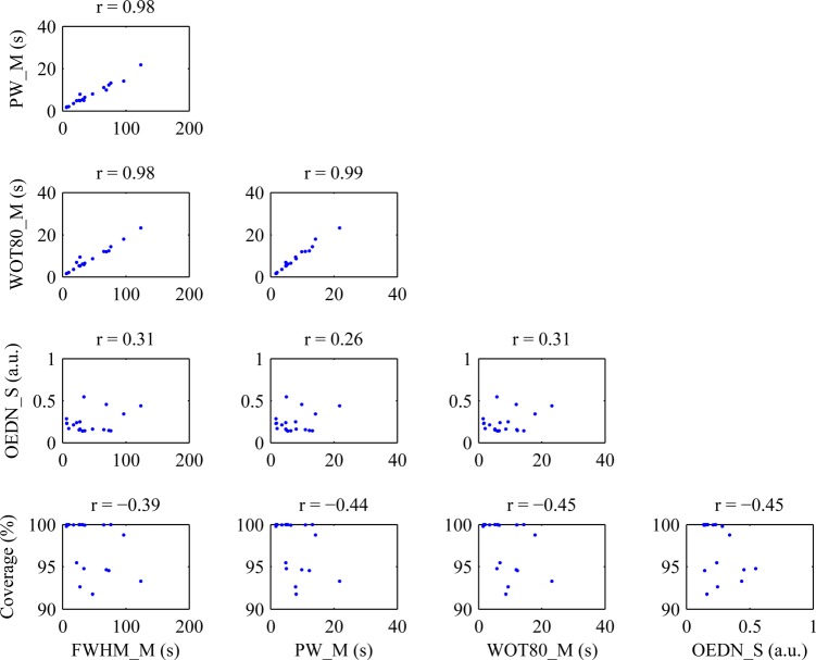
Contrast-enhanced ultrasound (CEUS) enables highly specific time-resolved imaging of vasculature by intravenous injection of ∼2 μm gas filled microbubbles. To develop a quantitative automated diagnosis of breast tumors with CEUS, breast tumors were induced in rats by administration of N-ethyl-N-nitrosourea. A bolus injection of microbubbles was administered and CEUS videos of each tumor were acquired for at least 3 min. The time-intensity curve of each pixel within a region of interest (ROI) was analyzed to measure kinetic parameters associated with the wash-in, peak enhancement, and wash-out phases of microbubble bolus injections since it was expected that the aberrant vascularity of malignant tumors will result in faster and more diverse perfusion kinetics versus those of benign lesions. Parameters were classified using linear discriminant analysis to differentiate between benign and malignant tumors and improve diagnostic accuracy. Preliminary results with a small dataset (10 tumors, 19 videos) show 100% accuracy with fivefold cross-validation testing using as few as two choice variables for training and validation. Several of the parameters which provided the best differentiation between malignant and benign tumors employed comparative analysis of all the pixels in the ROI including enhancement coverage, fractional enhancement coverage times, and the standard deviation of the envelope curve difference normalized to the mean of the peak frame. Analysis of combinations of five variables demonstrated that pixel-by-pixel analysis produced the most robust information for tumor diagnostics and achieved 5 times greater separation of benign and malignant cases than ROI-based analysis.
Figures
Similar articles
-
Prospective evaluation of contrast-enhanced ultrasound of breast BI-RADS 3-5 lesions.BMC Med Imaging. 2020 Jun 17;20(1):66. doi: 10.1186/s12880-020-00467-2. BMC Med Imaging. 2020. PMID: 32552678 Free PMC article.
-
Quantitative evaluation of contrast-enhanced ultrasound after intravenous administration of a microbubble contrast agent for differentiation of benign and malignant thyroid nodules: assessment of diagnostic accuracy.Eur Radiol. 2012 Jun;22(6):1357-65. doi: 10.1007/s00330-012-2385-6. Epub 2012 Feb 10. Eur Radiol. 2012. PMID: 22322310
-
Differentiation between benign and malignant breast tumors using kinetic features of real-time harmonic contrast-enhanced ultrasound.Acta Radiol. 2012 May 1;53(4):382-8. doi: 10.1258/ar.2012.110562. Epub 2012 Mar 20. Acta Radiol. 2012. PMID: 22434928
-
Quantitative evaluation of microvascular blood flow by contrast-enhanced ultrasound (CEUS).Clin Hemorheol Microcirc. 2011;49(1-4):137-49. doi: 10.3233/CH-2011-1464. Clin Hemorheol Microcirc. 2011. PMID: 22214685 Review.
-
Italian Society of Cardiovascular Echography (SIEC) Consensus Conference on the state of the art of contrast echocardiography.Ital Heart J. 2004 Apr;5(4):309-34. Ital Heart J. 2004. PMID: 15185894 Review.
Cited by
-
2-tier in-plane motion correction and out-of-plane motion filtering for contrast-enhanced ultrasound.Invest Radiol. 2014 Nov;49(11):707-19. doi: 10.1097/RLI.0000000000000074. Invest Radiol. 2014. PMID: 24901545 Free PMC article.
-
Quantification and Classification of Contrast Enhanced Ultrasound Breast Cancer Data: A Preliminary Study.Diagnostics (Basel). 2022 Feb 6;12(2):425. doi: 10.3390/diagnostics12020425. Diagnostics (Basel). 2022. PMID: 35204514 Free PMC article.
-
Longitudinal characterization of local perfusion of the rat placenta using contrast-enhanced ultrasound imaging.Interface Focus. 2019 Oct 6;9(5):20190024. doi: 10.1098/rsfs.2019.0024. Epub 2019 Aug 16. Interface Focus. 2019. PMID: 31485312 Free PMC article.
-
Hyposialylated IgG activates endothelial IgG receptor FcγRIIB to promote obesity-induced insulin resistance.J Clin Invest. 2018 Jan 2;128(1):309-322. doi: 10.1172/JCI89333. Epub 2017 Nov 27. J Clin Invest. 2018. PMID: 29202472 Free PMC article.
-
Pixel-based approach to assess contrast-enhanced ultrasound kinetics parameters for differential diagnosis of rheumatoid arthritis.J Med Imaging (Bellingham). 2015 Jul;2(3):034503. doi: 10.1117/1.JMI.2.3.034503. Epub 2015 Sep 11. J Med Imaging (Bellingham). 2015. PMID: 27014713 Free PMC article.
References
-
- American Cancer Society, Cancer Facts & Figures 2011 (American Cancer Society, Atlanta, 2011).
Grants and funding
LinkOut - more resources
Full Text Sources
Other Literature Sources