

Integrated processing of contrast pulse sequencing ultrasound imaging for enhanced active contrast of hollow gas filled silica nanoshells and microshells
- PMID: 23616935
- PMCID: PMC3463889
- DOI: 10.1116/1.3694835
Integrated processing of contrast pulse sequencing ultrasound imaging for enhanced active contrast of hollow gas filled silica nanoshells and microshells
Abstract
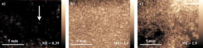
In recent years, there have been increasing developments in the field of contrast-enhanced ultrasound both in the creation of new contrast agents and in imaging modalities. These contrast agents have been employed to study tumor vasculature in order to improve cancer detection and diagnosis. An in vivo study is presented of ultrasound imaging of gas filled hollow silica microshells and nanoshells which have been delivered intraperitoneally to an IGROV-1 tumor bearing mouse. In contrast to microbubbles, this formulation of microshells provided strong ultrasound imaging signals by shell disruption and release of gas. Imaging of the microshells in an animal model was facilitated by novel image processing. Although the particle signal could be identified by eye under live imaging, high background obfuscated the particle signal in still images and near the borders of the tumor with live images. Image processing techniques were developed that employed the transient nature of the particle signal to selectively filter out the background signal. By applying image registration, high-pass, median, threshold, and motion filtering, a short video clip of the particle signal was compressed into a single image, thereby resolving the silica shells within the tumor. © 2012 American Vacuum Society.
Figures


Similar articles
-
Mechanically Tunable Hollow Silica Ultrathin Nanoshells for Ultrasound Contrast Agents.Adv Funct Mater. 2015 Jul 8;25(26):4049-4057. doi: 10.1002/adfm.201500610. Epub 2015 May 21. Adv Funct Mater. 2015. PMID: 26955300 Free PMC article.
-
Ultrasound Responsive Macrophase-Segregated Microcomposite Films for in Vivo Biosensing.ACS Appl Mater Interfaces. 2017 Jan 18;9(2):1719-1727. doi: 10.1021/acsami.6b10728. Epub 2017 Jan 5. ACS Appl Mater Interfaces. 2017. PMID: 28001041
-
Hollow silica and silica-boron nano/microparticles for contrast-enhanced ultrasound to detect small tumors.Biomaterials. 2012 Jul;33(20):5124-9. doi: 10.1016/j.biomaterials.2012.03.066. Epub 2012 Apr 11. Biomaterials. 2012. PMID: 22498299 Free PMC article.
-
Italian Society of Cardiovascular Echography (SIEC) Consensus Conference on the state of the art of contrast echocardiography.Ital Heart J. 2004 Apr;5(4):309-34. Ital Heart J. 2004. PMID: 15185894 Review.
-
Ultrasound contrast agents: basic principles.Eur J Radiol. 1998 May;27 Suppl 2:S157-60. doi: 10.1016/s0720-048x(98)00057-6. Eur J Radiol. 1998. PMID: 9652516 Review.
Cited by
-
Mechanically Tunable Hollow Silica Ultrathin Nanoshells for Ultrasound Contrast Agents.Adv Funct Mater. 2015 Jul 8;25(26):4049-4057. doi: 10.1002/adfm.201500610. Epub 2015 May 21. Adv Funct Mater. 2015. PMID: 26955300 Free PMC article.
-
Sol-gel synthesis and electrospraying of biodegradable (P2O5)55-(CaO)30-(Na2O)15 glass nanospheres as a transient contrast agent for ultrasound stem cell imaging.ACS Nano. 2015 Feb 24;9(2):1868-1877. doi: 10.1021/nn506789y. Epub 2015 Feb 3. ACS Nano. 2015. PMID: 25625373 Free PMC article.
-
Evolution of contrast agents for ultrasound imaging and ultrasound-mediated drug delivery.Front Pharmacol. 2015 Sep 15;6:197. doi: 10.3389/fphar.2015.00197. eCollection 2015. Front Pharmacol. 2015. PMID: 26441654 Free PMC article. Review.
-
Color Doppler ultrasound and gamma imaging of intratumorally injected 500 nm iron-silica nanoshells.ACS Nano. 2013 Jul 23;7(7):6367-77. doi: 10.1021/nn402507d. Epub 2013 Jul 1. ACS Nano. 2013. PMID: 23802554 Free PMC article.
-
Hollow iron-silica nanoshells for enhanced high intensity focused ultrasound.J Surg Res. 2014 Aug;190(2):391-8. doi: 10.1016/j.jss.2014.05.009. Epub 2014 May 10. J Surg Res. 2014. PMID: 24972734 Free PMC article.
References
Grants and funding
LinkOut - more resources
Full Text Sources
Other Literature Sources