

The potential for an enhanced role for MRI in radiation-therapy treatment planning
- PMID: 23617289
- PMCID: PMC4527434
- DOI: 10.7785/tcrt.2012.500342
The potential for an enhanced role for MRI in radiation-therapy treatment planning
Abstract
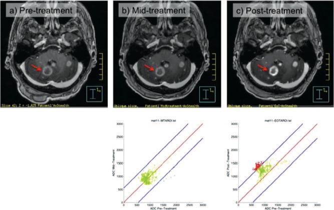
The exquisite soft-tissue contrast of magnetic resonance imaging (MRI) has meant that the technique is having an increasing role in contouring the gross tumor volume (GTV) and organs at risk (OAR) in radiation therapy treatment planning systems (TPS). MRI-planning scans from diagnostic MRI scanners are currently incorporated into the planning process by being registered to CT data. The soft-tissue data from the MRI provides target outline guidance and the CT provides a solid geometric and electron density map for accurate dose calculation on the TPS computer. There is increasing interest in MRI machine placement in radiotherapy clinics as an adjunct to CT simulators. Most vendors now offer 70 cm bores with flat couch inserts and specialised RF coil designs. We would refer to these devices as MR-simulators. There is also research into the future application of MR-simulators independent of CT and as in-room image-guidance devices. It is within the background of this increased interest in the utility of MRI in radiotherapy treatment planning that this paper is couched. The paper outlines publications that deal with standard MRI sequences used in current clinical practice. It then discusses the potential for using processed functional diffusion maps (fDM) derived from diffusion weighted image sequences in tracking tumor activity and tumor recurrence. Next, this paper reviews publications that describe the use of MRI in patient-management applications that may, in turn, be relevant to radiotherapy treatment planning. The review briefly discusses the concepts behind functional techniques such as dynamic contrast enhanced (DCE), diffusion-weighted (DW) MRI sequences and magnetic resonance spectroscopic imaging (MRSI). Significant applications of MR are discussed in terms of the following treatment sites: brain, head and neck, breast, lung, prostate and cervix. While not yet routine, the use of apparent diffusion coefficient (ADC) map analysis indicates an exciting future application for functional MRI. Although DW-MRI has not yet been routinely used in boost adaptive techniques, it is being assessed in cohort studies for sub-volume boosting in prostate tumors.
Figures
References
-
- Tubiana M. Can we reduce the incidence of second primary malignancies occurring after radiotherapy? A critical review. Radiother Oncol 91, 4–15 (2009). DOI: 10.1016/j.radonc.2008.12.016. - PubMed
-
- Desar I., Van Herpen C., Van Laarhoven H., Barentsz J., Oyen W., Van Der Graaf W. Beyond RECIST: molecular and functional imaging techniques for evaluation of response to targeted therapy. Cancer treatment reviews 35, 309–321 (2009). DOI: 10.1016/j.ctrv.2008.12.001. - PubMed
-
- Fallone B., Carlone M., Murray B., Rathee S., Stanescu T., Steciw S., Wachowicz K., Kirkby C. TU-C-M100F-01: development of a linac-MRI system for real-time ART. Medical Physics 34, 2547 (2007). DOI: 10.1118/1.2761342.
-
- Raaymakers B., Lagendijk J., Overweg J., Kok J., Raaijmakers A., Kerkhof E., van der Put R., Meijsing I., Crijns S., Benedosso F. Integrating a 1.5 T MRI scanner with a 6 MV accelerator: proof of concept. Physics in Medicine and Biology 54, N229 (2009). DOI: 10.1088/0031-9155/54/12/N01. - PubMed
-
- Oborn B., Metcalfe P., Butson M., Rosenfeld A., Keall P. Electron contamination modeling and skin dose in 6 MV longitudinal field MRIgRT: impact of the MRI and MRI fringe field. Medical Physics 39, 874 (2012). DOI: 10.1118/1.3676181. - PubMed
Publication types
MeSH terms
Substances
LinkOut - more resources
Full Text Sources
Other Literature Sources
Medical
