. 2013 Apr 24:13:14.
doi: 10.1186/1471-213X-13-14.

Development, organization, and remodeling of phoronid muscles from embryo to metamorphosis (Lophotrochozoa: Phoronida)

Affiliations

Development, organization, and remodeling of phoronid muscles from embryo to metamorphosis (Lophotrochozoa: Phoronida)

Elena N Temereva et al. BMC Dev Biol. .

Abstract

Background: The phoronid larva, which is called the actinotrocha, is one of the most remarkable planktotrophic larval types among marine invertebrates. Actinotrochs live in plankton for relatively long periods and undergo catastrophic metamorphosis, in which some parts of the larval body are consumed by the juvenile. The development and organization of the muscular system has never been described in detail for actinotrochs and for other stages in the phoronid life cycle.

Results: In Phoronopsis harmeri, muscular elements of the preoral lobe and the collar originate in the mid-gastrula stage from mesodermal cells, which have immigrated from the anterior wall of the archenteron. Muscles of the trunk originate from posterior mesoderm together with the trunk coelom. The organization of the muscular system in phoronid larvae of different species is very complex and consists of 14 groups of muscles. The telotroch constrictor, which holds the telotroch in the larval body during metamorphosis, is described for the first time. This unusual muscle is formed by apical myofilaments of the epidermal cells. Most larval muscles are formed by cells with cross-striated organization of myofibrils. During metamorphosis, most elements of the larval muscular system degenerate, but some of them remain and are integrated into the juvenile musculature.

Conclusion: Early steps of phoronid myogenesis reflect the peculiarities of the actinotroch larva: the muscle of the preoral lobe is the first muscle to appear, and it is important for food capture. The larval muscular system is organized in differently in different phoronid larvae, but always exhibits a complexity that probably results from the long pelagic life, planktotrophy, and catastrophic metamorphosis. Degeneration of the larval muscular system during phoronid metamorphosis occurs in two ways, i.e., by complete or by incomplete destruction of larval muscular elements. The organization and remodeling of the muscular system in phoronids exhibits the combination of protostome-like and deuterostome-like features. This combination, which has also been found in the organization of some other systems in phoronids, can be regarded as an important characteristic and one that probably reflects the basal position of phoronids within the Lophotrochozoa.


Figures

Figure 1
Figure 1
First steps of Phoronopsis harmeri myogenesis. In all images, the apical is to the top, and the ventral is to the right, except in B and C where the ventral side is toward the reader. A. Photograph of live mid-gastrula. B. Confocal image of the midgastrula stained with phalloidin; z-projection of 20 middle optical sections; frontal view. C. Mid-gastrula stained with phalloidin, 3D-projection of ventral side viewed from the dorsal side. The muscular cells (arrowheads) spread on the ventral wall of the embryo, above the archenteron (ar). D. Late gastrula with myoepithelial cells (mc), which have immigrated into the posterior part of the embryo and formed two lateral tiers (lt). E. Late gastrula stained with phalloidin, 3D-projection of first muscular system (blue) in the embryo (yellow); lateral view. F. Sagittal semi-thin section of late gastrula, in which hindgut begins to form as a posterior depression (pd) of the ectoderm. GJ. Ultrastructural organization of the first muscle cells, which have immigrated from the archenteron anterior wall; sagittal sections of the embryo. G. Basal processes (closed arrowheads) of muscle cells. H. First myofilaments (arrows) in the basal cytoplasm of muscle cells. I. The cilium with basal body (bb) and striated rootlet (sr) in the muscle cell. J. Part of the lining of the anterior pole of the late gastrula. The lining is formed by myoepithelial cells, which connect to each other via desmosomes (double arrowheads) and have a nucleus (n) with a nucleolus (nu). Abbreviations: ap – apical plate; bc – blastocoels; bp – blastopore; ep – epidermis.
Figure 2
Figure 2
Organization of the muscular system in early and young larva of Phoronopsis harmeri. In all photographs, animals are viewed from the lateral side; the ventral side is to the right, and apical side is to the top. A, C. Photographs of live larvae. B, D. 3D-reconstructions of muscular system (blue) of larvae stained with phalloidin (yellow). A, B. Early actinotrocha. C, D. Young larva. EH. 3D-reconstructions of muscular system of a young larva stained with phalloidin (EH) and with Hoechst (F). The position of the mouth is indicated by asterisk. E. Whole larva. F. Internal surface of the preoral lobe with radial muscles (rm) and their basal nuclei (arrowheads). G. Musculature of the edge of the preoral lobe includes annular muscle (am) and the bases of radial muscles. H. Esophageal musculature. Abbreviations: ap – apical plate; ar – archenteron; bc – blastocoels; es – esophageal musculature; lfc – laterofrontal cells are marked by long thick microvilli; t – lateral tiers of muscle cells; mc – muscle cells; pl – preoral lobe; plp – precursor of the preoral lobe; of – oral field; st – stomach; tc – transverse collar muscles; tg – tentacular ridge; tm – transversal muscles of the preoral lobe; vlm – ventro-lateral muscle.
Figure 3
Figure 3
Organization of the muscular system in different larval stages of Phoronopsis harmeri. In all photographs, the apical pole of larva is to the top. A. Live 6-day-old larva, lateral view, ventral side is to the right. BI. 3D-reconstructions of the muscular system of larvae stained with phalloidin. B. Whole 6-day-old larva viewed from the ventral side. C. Dorsal side of the base of the preoral lobe in 6-day-old larva. The chiasm of the radial muscles is indicated by arrowheads. D. A 9-day-old larva viewed from the ventral side. E. An 11-day-old larva viewed from the ventrolateral side. F. A 15-day-old larva, lateral view, ventral side is to the right. G. Anterior body part of a 15-day-old larva viewed from the dorsal side. The most dorsal optical sections have been excised. H. Anterior part of a 15-day-old larva viewed from the ventral side. The most ventral optical sections have been excised. Processes of the esophageal muscle cells, which contact the collar ring muscle, are shown by arrowheads. I. The trunk of a 24-day-old larva, ventral view. Abbreviations: am – annular muscle of the preoral lobe; cr – collar ring muscle; es – esophageal musculature; hd – hood depressors; lg – left group of radial muscles of the preoral lobe; mg – midgut; phd – precursor of the hood depressor; rg – right group of radial muscles of the preoral lobe; t – tentacle; tc – transverse collar muscles; te – tentacle elevator; tf – telotroch flexor; tg – tentacular ridge; tr – trunk retractor; vlm – ventrolateral muscles.
Figure 4
Figure 4
Organization of muscular system in advanced and competent larvae of Phoronopsis harmeri. In all photographs, the apical pole of the larva is to the top. A. Photograph of live larva, lateral view, the ventral side is to the right. BE. 3D-reconstructions of the muscular system of larvae stained with phalloidin. F – H. Z-projections of the muscular system of larvae stained with phalloidin. A. Competent larva. B. Overview of the musculature of a competent larva; lateral view. C. Musculature of anterior part of an advanced larva, lateral view. D. Collar region of an advanced larva with tentacular coelom (c2), collar ring muscle (cr), tentacle elevators (te), and tentacle depressors (td); lateral view, the ventral side is to the right. E. Apical body part of an advanced larva with apical plate (ap), paired hood depressors (hd), and esophageal musculature (es); dorsal view. F. Tentacular musculature of a competent larva. G. Overview of the musculature of a competent larva; lateral view, the ventral side is to the right. All main muscles are shown. H. Overview of the musculature of an advanced larva; lateral view, the ventral side is to the right. Abbreviations: bm – blood mass; es – esophageal musculature; eso – esophagus, lfc – laterofrontal cells of postoral ciliated band; ms – metasomal sack; st – stomach; t – tentacle; tt – telotroch.
Figure 5
Figure 5
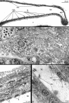
Organization of the hood musculature in Phoronopsis harmeri larvae. longitudinal semithin (A) and thin (B-D) sections. A. Anterior part of the hood. B. Annular muscle. C. The projection of the radial muscle cell. D. Organization of the muscle cell. Abbreviations: am – annular muscle; bc – blastocoel; cm – circular muscle; m – mitochondrion; mf – myofilaments; n – nucleus; nf – nerve fibers; rm – radial muscle; s – sarcomere; v – vestibulum; Z – Z-line. bb – basal body; c – cilium; G – Golgi apparatus.
Figure 6
Figure 6
Organization of the hood elevators and the esophageal musculature in Phoronopsis harmeri larvae. A. Fine morphology of hood elevators (arrows) (SEM). B. Cross section of hood elevator; muscle cells contact basal lamina via hemidesmosomes (open arrowheads). C. Semi-thin sagittal section of the esophagus (eso) with complete muscular lining (mc). D, E. Longitudinal fine section of muscular lining of the esophagus. D. Muscule cells contact basal lamina via hemidesmosomes (open arrowheads). E. Thick collagenous fibers between membranes of muscle cells are indicated by arrowheads (closed arrowheads). Abbreviations: bc – blastocoel; bl – basal lamina; ep – epidermis; mf – myofilaments; nf – nerve fibers.
Figure 7
Figure 7
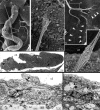
Hood depressors of Phoronopsis harmeri larva. A. Fine morphology of hood depressor (closed arrowheads), which passes along the wall of the collar to the circular ring muscle (cr). B. Semi-thin cross section of the central part of the preoral lobe and preoral coelom (c1) with upper portion of hood depressors (hd). C, D. Cross sections of the preoral lobe. C. Singular branch of the hood depressor is surrounded by thin projections of coelothelial cell (open arrowheads). D. Hood depressor muscle bunches (blue) are integrated between the coelothelial cells of preoral coelom (c1) lining. Abbreviations: bc – blastocoel; tc – transverse muscles of the collar.
Figure 8
Figure 8
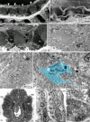
Fine morphology and ultrastructure of the collar ring muscle and tentacle elevators of Phoronopsis harmeri larvae. AC. Fine morphology of the collar ring muscle and tentacle elevators (SEM). A. Overview of the collar ring muscle (white arrowheads). B. Part of the collar ring muscle (cr) and two tentacle elevators (te) in a competent larva. C. Disorderly net of muscle cells of the collar ring muscle in an advanced larva. DF. Ultrastructure of the collar ring muscle (TEM). D. Overview of muscle cells with nueclei (n). E. Desmosomes (arrows) between muscle cells. F. Organization of one of the muscle cells (light blue), which has a cilium, basal body (bb), accessory centriole (ac), several Golgy complexes (G), and long processes that spread on the basal lamina (bl) and form bulbs (black arrowheads) opposite the nerve fibers (ne). G. Semi-thin cross section of the tentacle in a competent larva shows tentacular blood vessel (bv), coelomic lining (cl), frontal side epidermis (fs), the base of the tentacle elevator (teb), and the terminal bulge of the tentacle elevator (tet). H. Part of longitudinal section of the tentacle; a muscle cell with sarcomeres (s) is evident. I. muscle cells of the tentacle elevator (te), which is squeezed between the basal lamina (bl) of epidermis (ep) and the cells of the coelomic lining (cl) of the tentacular coelom.
Figure 9
Figure 9
Organization of trunk retractors, telotroch flexor, and telotroch constrictor in Phoronopsis harmeri larvae. A. Overview of two trunk retractors (tr) in the trunk coelom (SEM). B. Upper portion of the trunk retractor contacts the coelomic lining (cl) of the trunk coelom and collar ring muscle (cr) (SEM). C. Lower (posterior) portion of trunk retractor forms numerous long processes (white arrowheads) (SEM). D. Fine cross section of trunk retractor (TEM). Muscle cells connect to each other via desmosomes (black arrowheads). E. Middle portion of the trunk retractor contains nuclei of muscle cells. F. Cross section of larval trunk. Cells of the trunk coelom (c3) somatopleura form telotroch flexors and connect to each other via desmosomes (black arrowhead). G. Epidermal cells near the telotroch contain cross-striated-like myofilaments (closed arrowheads) in apical cytoplasm (TEM). Abbreviations: bc – blastocoel; bl – basal lamina; c3 – trunk coelom; mi – microvilli; ms – metasomal sack; n – nucleus.
Figure 10
Figure 10
Metacoel lining and blood vessels in Phoronopsis harmeri larvae. A-B: Longitudinal fine sections (TEM); longitudinal myofilaments are indicated by concave arrowheads; circular myofilaments are marked by straight arrowheads. C-D: Semi thin cross sections. A. Lining of the body wall. B. Lining of the digestive tract. C. The dorsal blood vessel (dv). D. The lateral blood vessel (lv). Abbreviations: bc – blastocoel; bl – basal lamina; cds – cells of the digestive system; c3 – trunk coelom; cl – cells of coelomic lining; st – stomach; vm – ventral mesentery.
Figure 11
Figure 11
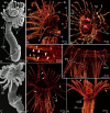
Metamorphosis of Phoronopsis harmeri. In all images, the apical is to the top. A. First stage of the metamorphosis (3 minutes after metamorphosis has begun) – animal with completely everted metasomal sack, which forms the juvenile body, is divided into the anterior trunk part (atp), the posterior trunk part (ptp), and the ampulla (a) (SEM). B. Head region of animal at the first step of metamorphosis, stained with phalloidin. 3D-reconstruction of muscular system; lateral view. C. Second step of the metamorphosis (10 minutes after metamorphosis has begun) – animal with engulfed preoral lobe (pl) (SEM). D. The same stage, 3D-reconstruction of musculature of animal head region stained with phalloidin; ventro-lateral view. E. Tentacles with degenerated (arrows) and complete (arrowheads) muscles. F. Oral view of newly formed juvenile 20 minutes after metamorphosis has begun. 3D-reconstruction of musculature of head region. Degenerated muscles are shown by arrows. G. The same stage; higher magnification of tentacles and lophophore with newly formed tentacle longitudinal muscles (tlm) and circular muscle of the tentacular coelom (mc2). H. Oral view of head region of 4-day-old juvenile. Degenerated muscles are shown by arrows. Abbreviations: am – annular muscle of the preoral lobe; ao – apical organ; cr – collar ring muscle; dt – definitive tentacles; es – esophageal musculature; fo – frontal organ; hd – hood depressor; lm – longitudinal muscles of the trunk; mt – metamorphic tentacles; tc – telotroch constructor; te – tentacle elevator; tf – telotroch flexor; tr – trunk retractor; tt – telotroch.
Figure 12
Figure 12
Organization of musculature in 4-day-old (A-C, E-F) and metamorphic (D) Phoronopsis harmeri. A. Borders with desmosome-like contacts (open arrowheads) between muscle cell (mc) of the tentacle elevator base and cell of the tentacular coelom lining (cl). B. Cross section of the tentacles with muscle cells of the tentacle elevator base, cells of the coelomic lining, and blood vessel (bv). Desmosomes between two muscle cells and between the muscle and coelothelial cells are indicated by opened arrowheads. C. Coelomocites include parts of degenerated muscular cells (double arrowheads). D. Longitudinal section of esophageal muscular lining with desmosomes (arrowheads) between cells. E. Coelomic lining (cl) of the trunk coelom (tc) and esophageal musculature (mc); longitudinal section of the esophagus (eso). F. Semi-thin cross section of the tentacle with blood vessel and degenerated muscles (dm) and erythrocyte (er) inside, base of the tentacle elevator (teb), coelomic cavity (c2) with coelomocites (cc), cells of the coelomic lining contain huge phagosomes (closed arrowheads).

Similar articles

Cited by

References

    1. Hejnol A, Obst M, Stamatakis A, Ott M, Rouse GW, Edgecombe GD, Martinez P, Baguñà J, Bailly X, Jondelius U, Wiens M, Müller WEG, Seaver E, Wheeler WC, Martindale MQ, Giribet G, Dunn CW. Assessing the root of bilaterian animals with scalable phylogenomic methods. Proc Biol Sci. 2009;276:4261–4270. doi: 10.1098/rspb.2009.0896. - DOI - PMC - PubMed
    1. Paps J, Baguñà J, Riutort M. Bilaterian Phylogeny: a broad sampling of 13 nuclear genes provides a new Lophotrochozoa phylogeny and supports a paraphyletic basal acoelomorpha. Mol Biol Evol. 2009;26(10):2397–2406. doi: 10.1093/molbev/msp150. - DOI - PubMed
    1. Cohen B, Weydmann A. Molecular evidence that phoronids are a subtaxon of brachiopods (Brachiopoda: Phoronata) and that genetic divergence of metazoan phyla began long before the early Cambrian. Organisms, Divers Evol. 2005;5:253–273. doi: 10.1016/j.ode.2004.12.002. - DOI
    1. Pennerstorfer M, Scholtz G. Early cleavage in Phoronis muelleri (Phoronida) displays spiral features. Evol Dev. 2012;14(6):484–500. doi: 10.1111/ede.12002. - DOI - PubMed
    1. Temereva EN, Malakhov VV. Embryogenesis in phoronids. Invert Zool. 2012;9(1):1–39.

Publication types