Review
. 2013 Apr 24;44(1):27.
doi: 10.1186/1297-9716-44-27.

Columnaris disease in fish: a review with emphasis on bacterium-host interactions

Affiliations
Review

Columnaris disease in fish: a review with emphasis on bacterium-host interactions

Annelies Maria Declercq et al. Vet Res. .

Abstract

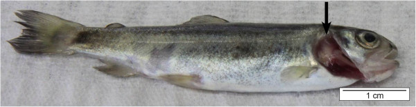
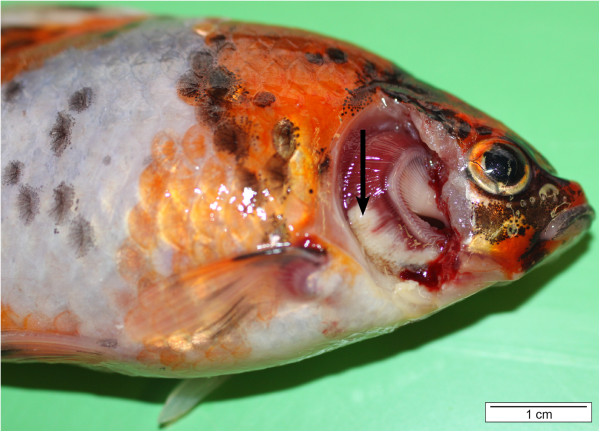
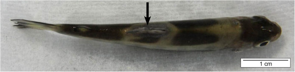
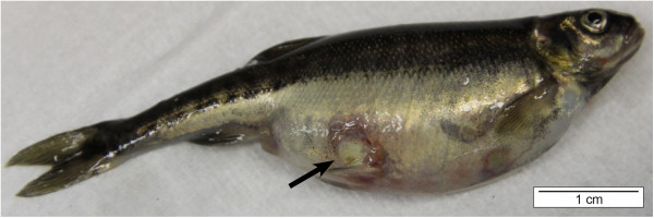
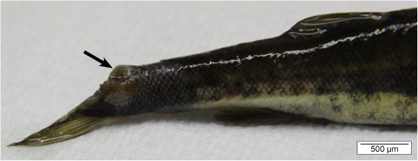
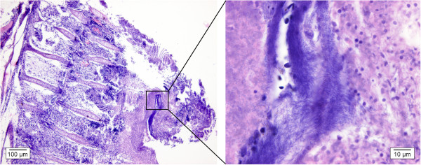
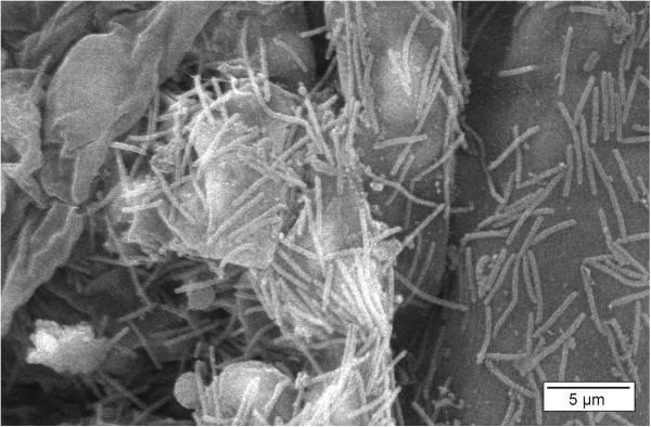
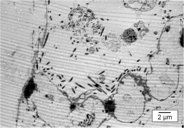
Flavobacterium columnare (F. columnare) is the causative agent of columnaris disease. This bacterium affects both cultured and wild freshwater fish including many susceptible commercially important fish species. F. columnare infections may result in skin lesions, fin erosion and gill necrosis, with a high degree of mortality, leading to severe economic losses. Especially in the last decade, various research groups have performed studies aimed at elucidating the pathogenesis of columnaris disease, leading to significant progress in defining the complex interactions between the organism and its host. Despite these efforts, the pathogenesis of columnaris disease hitherto largely remains unclear, compromising the further development of efficient curative and preventive measures to combat this disease. Besides elaborating on the agent and the disease it causes, this review aims to summarize these pathogenesis data emphasizing the areas meriting further investigation.


Figures

Figure 1
Figure 1
F. columnare elicited gill lesions in rainbow trout (Oncorhynchus mykiss) fry (operculum removed). In young fish, the disease is mostly acute and the gill is the major site of damage. The lesions are exhibited by pale necrotic areas (arrow, bar = 1 cm).
Figure 2
Figure 2
Gill lesions in a shubunkin (Carassius auratus) (operculum removed) caused by F. columnare. Yellowish-white areas of degeneration are visible in the ventral part of the first gill arch (arrow). When the F. columnare infection spreads rapidly throughout the gill lamellae, the fish may die in a short period of time without any other apparent lesions. Respiratory distress, caused by damage to the gills, appears to be the cause of death (bar = 1 cm).
Figure 3
Figure 3
F. columnare induced a saddleback lesion (arrow) in rainbow trout fry. The lesion is visible as a discoloration starting around its common location at the base of the dorsal fin and extending laterally to encircle the fish resembling a saddle. Hence, the descriptive term “saddle-back” is often used and the disease is denoted as “saddle-back disease” (bar = 1 cm).
Figure 4
Figure 4
Skin ulceration (arrow) in a minnow (Phoxinus phoxinus) caused by F. columnare. The lesion has progressed into deeper skin layers, exposing the musculature. The edge of the ulceration displays a distinct reddish tinge and its centre is covered with yellowish-white mucus (bar = 1 cm).
Figure 5
Figure 5
Tail fin erosion in a minnow caused by F. columnare. Columnaris disease led to complete disappearance of the upper half of the fin and exposing the underlying musculature (arrow, bar = 500 μm).
Figure 6
Figure 6
Gill section of a koi carp infested with F. columnare. In the left figure, extensive loss of branchial structures is visible. This is an advanced stage of the disease in which the filaments and the lamellae have fused and the gill epithelium is destroyed (H&E, bar = 100 μm). Complete clubbing of gill filaments may finally result in circulatory failure and extensive internal hemorrhage. A detail of this is depicted in the right figure, where F. columnare bacteria are visible as long, slender, purplish structures in between remnants of the gill tissue (H&E, bar = 10 μm).
Figure 7
Figure 7
Gill of a rainbow trout infested with F. columnare. This scanning electron microscopic (SEM) picture of an affected gill arch reveals the presence of long, thin, rod-shaped bacterial cells, approximately 0.3-0.5 μm wide and 3-10 μm long (SEM, bar = 5 μm).
Figure 8
Figure 8
Gill lamellae of a koi carp following infection by F. columnare. This transmission electron microscopic picture clearly demonstrates the loss of structure of the gill lamellae. The F. columnare bacteria are lining up around the remnants of these lamellae, and are closely associated with the gill epithelium (TEM, bar = 2 μm).

Similar articles

Cited by

References

    1. Bernardet JF, Bowman JP. In: The Prokaryotes: A Handbook on the Biology of Bacteria: Volume 7: Proteobacteria: Delta and Epsilon Subclasses. Deeply Rooting Bacteria. Dworkin M, Falkow S, editor. New York: Springer Science+Business Media, LLC; 2006. The genus Flavobacterium; pp. 481–531.
    1. Bernardet JF, Nakagawa Y, Holmes B. Proposed minimal standards for describing new taxa of the family Flavobacteriaceae and emended description of the family. Int J Syst Evol Microbiol. 2002;52:1049–1070. doi: 10.1099/ijs.0.02136-0. - DOI - PubMed
    1. Bernardet JF, Segers P, Vancanneyt M, Berthe M, Kersters K, Vandamme P. Cutting a Gordian knot: emended classification and description of the genus Flavobacterium, emended description of the family Flavobacteriaceae, and proposal of Flavobacterium hydatis nom. nov. (basonym, Cytophaga aquatilis Strohl and Tait 1978) Int J Syst Evol Bacteriol. 1996;46:128–148. doi: 10.1099/00207713-46-1-128. - DOI
    1. Davis HS. A new bacterial disease in freshwater fishes. United States Bureau of Fisheries Bulletin. 1922;38:37–63.
    1. Ordal EJ, Rucker RR. Pathogenic myxobacteria. PSEBM. 1944;56:15–18.

Publication types

MeSH terms

LinkOut - more resources