Review
. 2013 Aug;15(8):1313-22.
doi: 10.1111/cmi.12152. Epub 2013 May 13.

Caenorhabditis elegans as a model for intracellular pathogen infection

Affiliations
Review

Caenorhabditis elegans as a model for intracellular pathogen infection

Keir M Balla et al. Cell Microbiol. 2013 Aug.

Abstract

The genetically tractable nematode Caenorhabditis elegans is a convenient host for studies of pathogen infection. With the recent identification of two types of natural intracellular pathogens of C. elegans, this host now provides the opportunity to examine interactions and defence against intracellular pathogens in a whole-animal model for infection. C. elegans is the natural host for a genus of microsporidia, which comprise a phylum of fungal-related pathogens of widespread importance for agriculture and medicine. More recently, C. elegans has been shown to be a natural host for viruses related to the Nodaviridae family. Both microsporidian and viral pathogens infect the C. elegans intestine, which is composed of cells that share striking similarities to human intestinal epithelial cells. Because C. elegans nematodes are transparent, these infections provide a unique opportunity to visualize differentiated intestinal cells in vivo during the course of intracellular infection. Together, these two natural pathogens of C. elegans provide powerful systems in which to study microbial pathogenesis and host responses to intracellular infection.


Figures

Fig. 1
Fig. 1
Features of the C. elegans intestine. A. Simplified body plan of C. elegans. Microbes are ingested by the pharynx (yellow), which helps degrade food before it reaches the intestine (purple). B. Close-up model of the field highlighted by the dotted box in (A). Divisions between intestinal cells are shown, as well as nuclei and the lumen. C. Cell biology of intestinal cells. D. Electron micrograph of a C. elegans intestinal cell.
Fig. 2
Fig. 2
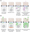
Intracellular pathogens studied in C. elegans and the cell biology of natural intracellular infections in C. elegans intestinal cells. A. Model of the predicted Orsay virus lifecycle and intestinal phenotypes associated with infection. Virions are drawn as purple hexagons, viral RNA is drawn as purple curved lines for positive strand transcripts and green for negative strand transcripts. Shorter lines represent small RNAs. Actin is drawn as vertical orange lines, terminal web as blue lines, and apical junctions as black ovals. B. Model of the N. parisii lifecycle and intestinal phenotypes associated with microsporidia infection. Spores are drawn as green ovals, polar tube is green line from invading spore, replicating meronts are irregular green shapes. Actin is drawn as vertical orange lines, terminal web as blue lines, and apical junctions as black ovals.

References

    1. Aballay A, Yorgey P, Ausubel FM. Salmonella typhimurium proliferates and establishes a persistent infection in the intestine of Caenorhabditis elegans. Curr Biol. 2000;10:1539–1542. - PubMed
    1. Andersen EC, Gerke JP, Shapiro JA, Crissman JR, Ghosh R, Bloom JS, et al. Chromosome-scale selective sweeps shape Caenorhabditis elegans genomic diversity. Nat Genet. 2012;44:285–290. - PMC - PubMed
    1. Brenner S. The genetics of Caenorhabditis elegans. Genetics. 1974;77:71–94. - PMC - PubMed
    1. Burton EA, Pendergast AM, Aballay A. The Caenorhabditis elegans ABL-1 tyrosine kinase is required for Shigella flexneri pathogenesis. Appl Environ Microbiol. 2006;72:5043–5051. - PMC - PubMed
    1. Casadevall A. Evolution of intracellular pathogens. Annu Rev Microbiol. 2008;62:19–33. - PubMed

Publication types

MeSH terms