

Review
doi: 10.1021/cr300384w.
Epub 2013 Apr 25.
Gates of enzymes
Affiliations
- PMID: 23617803
- PMCID: PMC3744840
- DOI: 10.1021/cr300384w
Item in Clipboard
Review
Gates of enzymes
Chem Rev.
.
No abstract available
Figures
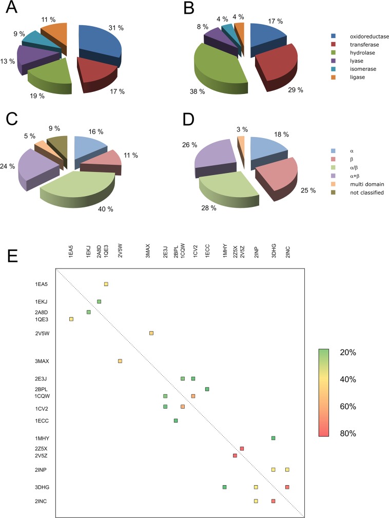
Distribution of (A) reviewed proteins and (B) proteins
from the
PDB database according to EC classes, (C) reviewed proteins and (D)
proteins from the PDB database according to SCOP classification, and
(E) identity matrix of reviewed enzymes (only the enzymes with sequence
identities above 20% are shown for clarity).
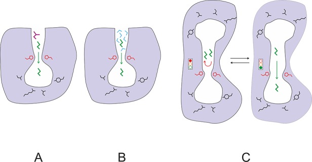
Schematic illustration of the molecular functions of protein gates:
(A) control of substrate access, (B) control of solvent access, (C)
control and synchronization of reactions. Protein is represented by
the area colored in gray, active site cavity by the area in white,
gating residues by red lines, substrate molecules by green or violet
lines, and water molecules by blue lines.
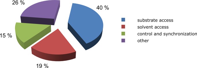
Distribution of gate functions; 71 proteins with 129 different
gates were analyzed. Percentages shown in the figure are based on
all of the identified functions of each gate; individual gate may
perform multiple functions. Detailed description of the analyzed proteins
is provided in Table 3.
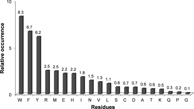
Relative occurrences of specific amino acid residues in
wing and
swinging door gates; 71 proteins with 129 gates were analyzed, and
154 residues that form wings or swinging doors were identified. Detailed
description of the analyzed proteins is provided in Table 3. Values were normalized against the frequency with
which each amino acid appears in all of the protein structures of
the UniProtKB/Swiss-Prot database (2012_07).
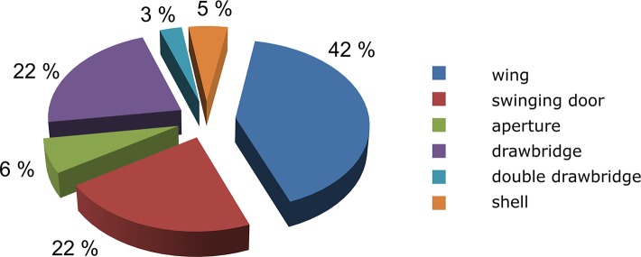
Frequencies
of different gate types based on analysis of 71 proteins
with 129 gates. Detailed description of the analyzed proteins is provided
in Table 3.
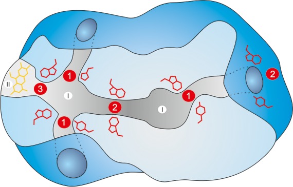
Locations of gates within
a protein structure. Schematic representation
of an enzyme with two active sites connected by a tunnel (I), a cofactor
cavity (II), and multiple access tunnels. Gating residues in red may
be located at the entrance to the active site (1), at the entrance
or the bottleneck of the tunnel (2), and between the active site cavity
and the cofactor cavity (3).
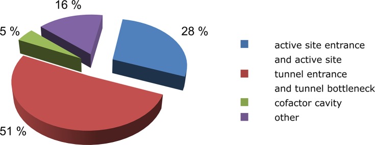
Distribution of gate locations within
protein structures based
on analysis of 71 proteins with 129 gates. Detailed description of
the analyzed proteins is provided in Table 3.
References
-
- Röthlisberger D.; Khersonsky O.; Wollacott A. M.; Jiang L.; DeChancie J.; Betker J.; Gallaher J. L.; Althoff E. A.; Zanghellini A.; Dym O.; Albeck S.; Houk K. N.; Tawfik D. S.; Baker D. Nature 2008, 453, 190. - PubMed
Publication types
MeSH terms
Substances
LinkOut - more resources
Full Text Sources
Other Literature Sources
