. 2013 Apr 23:10:128.
doi: 10.1186/1743-422X-10-128.

Influenza A virus induced bacterial otitis media is independent of virus tropism for α2,6-linked sialic acid

Affiliations

Influenza A virus induced bacterial otitis media is independent of virus tropism for α2,6-linked sialic acid

Kirsty R Short et al. Virol J. .

Abstract

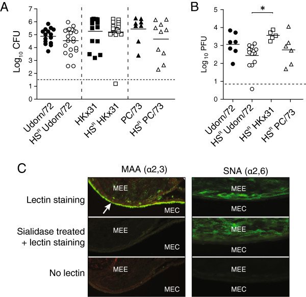
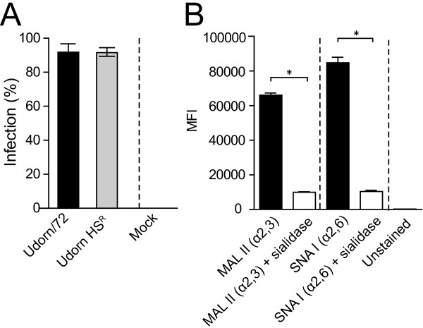
Background: Otitis media (OM) affects ≥80% of children before the age of three. OM can arise following co-infection with influenza A virus (IAV) and the bacterium Streptococcus pneumoniae. We have previously shown that H3 IAV strains (such as Udorn/72) induced a higher rate of bacterial OM than H1 strains (such as PR8/34). This was associated with more efficient replication of H3 strains in the middle ear.

Findings: Here, we assess if the increased replication of IAV strains such as Udorn/72 in the middle ear is dependent upon the binding of the viral HA to α2,6-linked sialic acid. Using murine and in vitro models, the present study shows that recognition of α2,6-linked sialic acid was not required to facilitate bacterial OM.

Conclusions: Taken together, these data suggest that other features of the HA mediate bacterial OM.


Figures

Figure 1
Figure 1
α2,6-linked sialic acid recognition does not affect the development of pneumococcal OM. A. Titres of S. pneumoniae EF3030lux in the middle ears of mice six-days after i.n. infection with wild-type or horse-serum resistant (HSR) IAV strains. Bacterial counts are represented as the average titre derived from the left and right ear of each mouse. Data is pooled from a minimum of two independent experiments. Statistical significance was determined using a Mann–Whitney U-Test between the relevant wild-type or HSR strain. Dashed line indicates the detection limit of the assay. B. Titres of IAV in the middle ears of mice six-days after i.n. infection with wild-type or horse-serum resistant (HSR) IAV strains. Viral tires are represented as the average titre derived from the left and right ear of each mouse. Data is pooled from a minimum of two independent experiments. Statistical significance was determined using a One-Way ANOVA with a Bonferroni post-correction and is denoted by an asterisk (p < 0.05). Dashed line indicates the detection limit of the assay. C. Sialic acid expression in the murine middle ear. Sections were labeled with Maackia amurensis agglutinin (MAA; α2,3-linked sialic acid) or Sambucus nigra agglutinin (SNA; α2,6-linked sialic acid) with or without pre-treatment with a sialidase. Positive staining is shown by an arrow. MEE: Middle Ear Epithelium; MEC: Middle Ear Cavity.
Figure 2
Figure 2
α2,6-linked sialic acid recognition does not affect infection of human middle ear epithelial cells (HMEECs). A. Percentage of HMEECs infected by wild-type Udorn/72 or horse serum resistant (HSR) Udorn/72 eight hours post-infection at an MOI of 1. Statistical significance was determined using a Mann–Whitney U-Test. Data represents mean ± SEM. B. HMEECs express α2,3- and α2,6-linked sialic acid. HMEECs were incubated with PBS containing 1% BSA (unstained), biotinylated MAL II (to detect α2,3-linked sialic acid), biotinylated SNA I (to detect α2,6-linked sialic acid) or biotinylated MAL II or SNA I following sialidase treatment (sialidase treated). Cells were subsequently stained with streptavidin-conjugated APC and measured by flow cytometry. The mean fluorescent intensity (MFI) is shown. Data is pooled from two independent experiments where each experiment was performed in duplicate. Data represents mean ± SEM. Statistical significance was determined using a Mann–Whitney U-Test and is denoted by one asterisk (p < 0.05).

References

    1. Massa HM, Cripps AW, Lehmann D. Otitis media: viruses, bacteria, biofilms and vaccines. Med J Aus. 2010;191:S44–S49. - PMC - PubMed
    1. Peltola VT, Boyd KL, McAuley JL, Rehg JE, McCullers JA. Bacterial sinusitis and otitis media following influenza virus infection in ferrets. Infect Immun. 2006;74:2562–2567. doi: 10.1128/IAI.74.5.2562-2567.2006. - DOI - PMC - PubMed
    1. Giebink GS, Berzins IK, Marker SC, Schiffman G. Experimental otitis media after nasal inoculation of streptococcus pneumoniae and influenza a virus in chinchillas. Infect Immun. 1980;30:445–450. - PMC - PubMed
    1. Short KR, Diavatopoulos DA, Thorton R. Influenza virus induces bacterial and non-bacterial otitis media. J Infect Dis. 2011;204:1857–1865. doi: 10.1093/infdis/jir618. - DOI - PMC - PubMed
    1. Short KR, Reading PC, Brown LE. Influenza-induced inflammation drives pneumococcal otitis media. Infect Immun. 2012;81(3):645–652. - PMC - PubMed

Publication types

MeSH terms

Substances