Paraneoplastic necrotizing myopathy associated with adenocarcinoma of the lung - a rare entity with atypical onset: a case report
- PMID: 23618006
- PMCID: PMC3641969
- DOI: 10.1186/1752-1947-7-112
Paraneoplastic necrotizing myopathy associated with adenocarcinoma of the lung - a rare entity with atypical onset: a case report
Abstract
Introduction: Inflammatory myopathies (such as dermatomyositis and polymyositis) are well-recognized paraneoplastic syndromes. However, paraneoplastic necrotizing myopathy is a more recently defined clinical entity, characterized by rapidly progressive, symmetrical, predominantly proximal muscle weakness with severe disability, and associated with a marked increase in serum muscle enzyme levels. Paraneoplastic necrotizing myopathy requires muscle biopsy for diagnosis, which typically shows massive necrosis of muscle fibers with limited or absent inflammatory infiltrates.
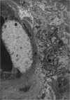
Case presentation: We report the case of an 82-year-old Italian-born Caucasian man who was admitted to hospital because of heart failure and two drop attacks. Over the following days, he developed progressive severe weakness, dysphagia, and dysphonia. Testing showed increasing serum muscle enzyme levels. Electromyography showed irritative myopathy of the proximal muscles and sensorimotor polyneuropathy. Muscle biopsy (left vastus lateralis) showed massive necrosis of muscle fibers with negligible inflammatory infiltrates, complement membrane attack complex deposition on endomysial capillaries, and moderate upregulation of major histocompatibility complex-I. Computed tomography of the thorax showed a nodular mass in the apex of the right lung. The patient was diagnosed with paraneoplastic necrotizing myopathy. In spite of high-dose corticoid therapy, he died 1 month later because of his aggressive cancer. Subsequent electron microscopic examination of a muscle biopsy specimen showed thickened walls and typical pipestem changes of the endomysial capillaries, with swollen endothelial cells. Poorly differentiated adenocarcinoma of the lung was confirmed on post-mortem histological examination.
Conclusions: Paraneoplastic necrotizing myopathy is a rare syndrome with outcomes ranging from fast progression to complete recovery. Treatment with corticosteroids is often ineffective, and prognosis depends mainly on the characteristics of the underlying cancer. This case shows that paraneoplastic necrotizing myopathy may have an atypical appearance, and should be considered in elderly patients with neoplastic disease. In this case, the diagnosis was delayed by the unusual clinical picture that suggested heart disease rather than muscle disease.
Figures


Similar articles
-
Paraneoplastic necrotizing myopathy: clinical and pathological features.Neurology. 1998 Mar;50(3):764-7. doi: 10.1212/wnl.50.3.764. Neurology. 1998. PMID: 9521271 Review.
-
Paraneoplastic necrotizing myopathy in a woman with breast cancer: a case report.J Med Case Rep. 2009 Nov 2;3:95. doi: 10.1186/1752-1947-3-95. J Med Case Rep. 2009. PMID: 19946512 Free PMC article.
-
Paraneoplastic Necrotizing Myopathy with a Mild Inflammatory Component: A Case Report and Review of the Literature.Case Rep Oncol. 2010 Apr 8;3(1):88-92. doi: 10.1159/000308714. Case Rep Oncol. 2010. PMID: 20740165 Free PMC article.
-
Prominent Asymmetric Muscle Weakness and Atrophy in Seronegative Immune-Mediated Necrotizing Myopathy.Diagnostics (Basel). 2021 Nov 8;11(11):2064. doi: 10.3390/diagnostics11112064. Diagnostics (Basel). 2021. PMID: 34829411 Free PMC article.
-
[Acquired necrotizing myopathies].Rev Med Interne. 2013 Jun;34(6):363-8. doi: 10.1016/j.revmed.2012.07.018. Epub 2012 Sep 19. Rev Med Interne. 2013. PMID: 22998975 Review. French.
Cited by
-
Paraneoplastic syndromes associated with lung cancer.World J Clin Oncol. 2014 Aug 10;5(3):197-223. doi: 10.5306/wjco.v5.i3.197. World J Clin Oncol. 2014. PMID: 25114839 Free PMC article. Review.
-
Muscle weakness and myalgia as the initial presentation of serous ovarian carcinoma: a case report.J Ovarian Res. 2014 Apr 23;7:43. doi: 10.1186/1757-2215-7-43. eCollection 2014. J Ovarian Res. 2014. PMID: 24812576 Free PMC article.
-
Rare case of Sjögren's syndrome antibody (SSA-Ro52kDA)-associated interstitial lung disease and myositis with pipe stem capillaries.BMJ Case Rep. 2021 Oct 7;14(10):e244133. doi: 10.1136/bcr-2021-244133. BMJ Case Rep. 2021. PMID: 34620633 Free PMC article.
-
Signal Recognition Particle in Human Diseases.Front Genet. 2022 Jun 8;13:898083. doi: 10.3389/fgene.2022.898083. eCollection 2022. Front Genet. 2022. PMID: 35754847 Free PMC article.
-
Necrotizing Autoimmune Myopathy: Clinicopathologic Study from a Single Tertiary Care Centre.Ann Indian Acad Neurol. 2018 Jan-Mar;21(1):62-67. doi: 10.4103/aian.AIAN_389_17. Ann Indian Acad Neurol. 2018. PMID: 29720800 Free PMC article.
References
-
- Shimizu J. [Malignancy-associated myositis] [Article in Japanese] Brain Nerve. 2010;62:427–432. - PubMed
LinkOut - more resources
Full Text Sources
Other Literature Sources
Miscellaneous

