. 2013 Apr 26:8:8.
doi: 10.1186/1749-8104-8-8.

Temporal and spatial requirements of Smoothened in ventral midbrain neuronal development

Affiliations

Temporal and spatial requirements of Smoothened in ventral midbrain neuronal development

Mianzhi Tang et al. Neural Dev. .

Abstract

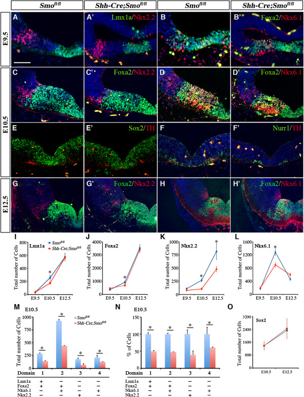
Background: Several studies have indicated that Sonic hedgehog (Shh) regulates the expansion of dopaminergic (DA) progenitors and the subsequent generation of mature DA neurons. This prevailing view has been based primarily on in vitro culture results, and the exact in vivo function of Shh signaling in the patterning and neurogenesis of the ventral midbrain (vMB) remains unclear.

Methods: We characterized the transcriptional codes for the vMB progenitor domains, and correlated them with the expression patterns of Shh signaling effectors, including Shh, Smoothened, Patched, Gli1, Gli2 and Gli3.

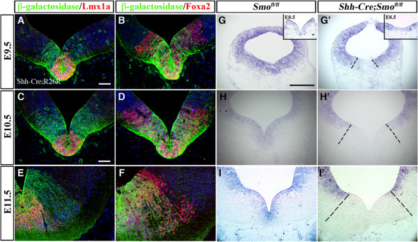
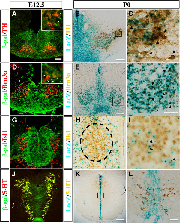
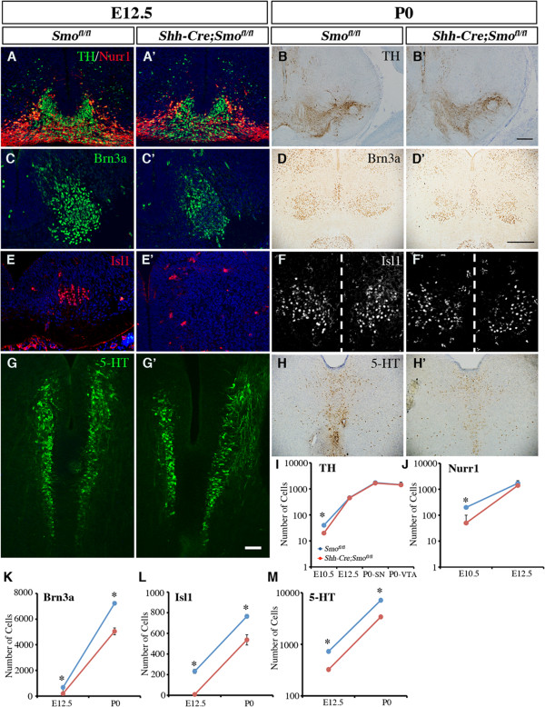
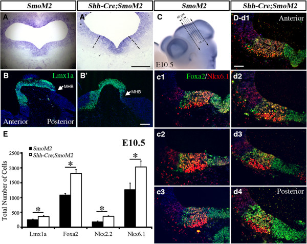
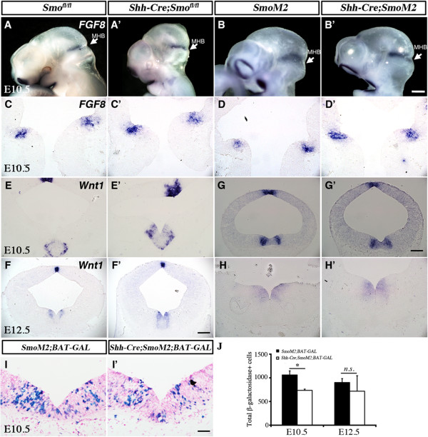
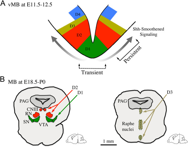
Results: While Shh and its downstream effectors showed robust expression in the neurogenic niche for DA progenitors at embryonic day (E)8 to E8.5, their expression shifted to the lateral domains from E9.5 to E12.5. Consistent with this dynamic change, conditional mutants with region-specific removal of the Shh receptor Smoothened in the vMB progenitors (Shh-Cre;Smo(fl/fl)) showed a transient reduction in DA progenitors and DA neurons at E10.5, but had more profound defects in neurons derived from the more lateral domains, including those in the red nucleus, oculomotor nucleus, and raphe nuclei. Conversely, constitutive activation of Smoothened signaling in vMB (Shh-Cre;SmoM2) showed transient expansion of the same progenitor population. To further characterize the nature of Shh-Smoothened signaling in vMB, we examined the BAT-GAL reporter and the expression of Wnt1 in vMB, and found that the antagonistic effects of Shh and Wnt signaling critically regulate the development of DA progenitors and DA neurons.

Conclusion: These results highlight previously unrecognized effects of Shh-Smoothened signaling in the region-specific neurogenesis within the vMB.


Figures

Figure 1
Figure 1
Distinct progenitor domains in the developing ventral midbrain (vMB) defined by a combinatorial code of transcription factors. (A-D) Double immunostaining for (A,C) Foxa2/Nkx6.1, (B,D) Lmx1a/Nkx2.2 and (inset) Foxa2/Nkx2.2 in vMB at embryonic day (E)8.5 (nine somites) and E9.5. (A) At E8.5, Foxa2 and Foxa2/Nkx6.1 defines domain D1 and D2. (B) Note that Lmx1a and Nkx2.2 are not expressed at E8.5. (C,D) At E9.5, Lmx1a/Foxa2, Foxa2/Nkx6.1, and Nkx2.2 define domains D1 to D3, respectively. (E-J) Confocal images show the expression pattern of (E,I) Foxa2/Nkx2.2, (F,J) Foxa2/Nkx6.1, (G) Foxa2/Lmx1a and (H) Olig2/Pax6 in vMB at E10.5 and E12.5. At both stages, Lmx1a/Foxa2, Foxa2/Nkx6.1, Nkx2.2-only and Nkx6.1-only define domains D1 to D4, respectively. Unlike in the ventral spinal cord, Olig2 and Pax6 were not detected in vMB at E10.5 Scale bars: (H) 50 μm, applied to A-H; (J) 100 μm, applied to I-J. (K) Schematic diagrams illustrating the D1 to D4 progenitor domains in vMB defined by a combinatorial code of transcription factors from E8.5 to E12.5.
Figure 2
Figure 2
Dynamic expression patterns of Shh signaling components in the developing ventral midbrain (vMB). (A) Immunohistochemical staining and in situ hybridization (insets) revealed Shh expression in the midline of vMB at embryonic day (E)8.5 (10 somites). (B-C and insets) Shh expression in vMB extended laterally from E9.5 toE10.5, then (D, inset) became restricted laterally at E11.5. (E-H) In situ hybridization for Smoothened from E8 to E11.5 show that Smoothened was diffusely expressed both dorsally and ventrally in midbrain and its expression became progressively restricted to the ventricular zone in vMB at E10.5 to E11.5. (F, G, H) Dashed lines outline the pia side of neurotube. (I-X) In situ hybridization for (I-L) Patched, (M-P) Gli1, (Q-T) Gli2, and (U-X) Gli3 from E8 to E11.5. All were expressed medially at E8 to E8.5 (7–10 somites), and then shifted laterally from E9.5 onwards. Scale bars: (A), 50 μm, applied to A, B; (C) 200 μm, applied to C, D, H; (E) 25 μm, applied to E, I M, Q, U; (F) 200 μm, applied to F, J, N, R, V; and (G) 500 μm, applied to G, K, O, S, W, L, P, T, X.
Figure 3
Figure 3
Spatial distribution of Sonic hedgehog (Shh) signaling effectors in ventral midbrain progenitor domains. (A-C) Double immunofluorescence staining of Shh and Nkx6.1 reveal that Shh proteins were expressed mainly in (A) the D1 region at embryonic day (E)9.5,(B) D1 and D2 regions at E10.5, and (C) D2 region at E11.5. (D-F) Double immunofluorescence staining of Shh and Nkx2.2 show partial colocalization from E9.5 to E10.5, but distinct separation at E11.5. (G-O) Combined in situ hybridization of Patched and Gli1 with immunohistochemistry for Nkx6.1 and Nkx2.2 show that Patched and Gli1 mRNA were expressed in D2 to D4 domains, especially enriched in the D3 region. Scale bars: (A) 25 μm, applied to A, D; (B) 50 μm, applied to B-C and E-F; (G) 100 μm, applied to G-O.
Figure 4
Figure 4
Region-specific removal of Smoothened in the ventral midbrain of Shh-Cre;Smofl/flmutants. (A-F) Using the R26R reporter line, we found that Shh-Cre drives recombination in D1 domain and the majority of D2 domain from embryonic day (E)9.5 to E11.5. (G-I’) In situ hybridization of Smoothened indicates complete removal of Smoothened in Shh-Cre;Smofl/fl mice from E9.5 to E11.5, and (G-G’ and insets) incomplete removal at E8.5 (eight somites). Scale bars: (A) 25 μm, applied to A-B; (C) 50 μm, applied to C-F; (G) 200 μm, applied to G and I’.
Figure 5
Figure 5
Transient reduction of dopaminergic (DA) progenitors in Shh-Cre;Smofl/flmutants. (A-B’) At embryonic day (E)9.5, Shh-Cre;Smofl/fl mutants showed no detectable reduction in progenitors expressing (A’) Lmx1a/Nkx2.2, or (B’) Foxa2/Nkx6.1. (C-F’) By contrast, Shh-Cre;Smofl/fl mutants showed a significant reduction in (C-C’, D-D’) Foxa2+, (C-C’) Nkx2.2+, (D-D’) Nkx6.1+, and (F-F’) Nurr1+ progenitors at E10.5, whereas (E-E’) Sox2+ progenitors were unchanged. (E-F’) Note that the DA neurons were also reduced at E10.5. (G’,H’) Foxa2+ and (H’) Nkx6.1+ progenitors from E12.5 Shh-Cre;Smofl/fl mutants showed no change compared with (G,H) controls, whereas (G’) the reduction in Nkx2.2+ progenitors persisted at E12.5. Scale bars: (A) 50 μm (applied to A to H’). (I-L) Quantification of total number of (I) Lmx1a+, (J) Foxa2+, (K) Nkx2.2+ and (L) Nkx6.1+ progenitors confirmed their transient reduction at E10.5. Student’s t test, n = 3 or 4. (M-N) Quantification of number of cells in each progenitor domains at E10.5 showed a consistent reduction from D1 to D4 domains at E10.5.
Figure 6
Figure 6
Fate mapping of dopaminergic neurons, red nucleus neurons, oculomotor neurons, and serotonergic neurons from Sonic hedgehog (Shh)-expressing cells. Using the Shh-Cre;R26R reporter line, we found that (A) tyrosine hydroxylase (TH)+ DA neurons, (D) Brn3a+ neurons in the red nucleus, (G) Isl1+ oculomoter neurons (H) and 5-hydroxytryptamine (5-HT)+ serotonergic neurons showed extensive coexpression of β-galactosidase at embryonic day (E)12.5. (A and D insets) Arrowheads show the colocalization of (A inset) TH (red) and (D inset) Brn3a (red) with β-galactosidase (green) at E12.5. At post-natal day (P)0, (B) TH+, (E) Brn3a+, (H) Isl1+ and (K) 5-HT+ neurons expressed within the LacZ-expressed regions (blue). (C,F,I,L) Higher magnifications of the boxed regions in (B,E,H,K), respectively. Black arrowheads indicate the colocalized cells. Scale bars: (A) 50 μm, applied to A, D, G, E); (B, F, H) 100 μm; (C) 50 μm; (E) 200 μm.
Figure 7
Figure 7
Persistent loss of neurons in the red nucleus, oculomotor nucleus and the serotonergic neurons, but not dopaminergic neurons, in Shh-Cre;Smofl/flmutants at embryonic day (E)12.5 to post-natal day (P)0. (A-B’) Nurr1 and tyrosine hydroxylase (TH) staining from E12.5 and P0 Shh-Cre;Smofl/fl mutants showed no change compared with controls. (C-D’) Brn3a, (E-F’) Islet1 and (G-H’) 5-Hydroxytryptamine (5-HT) revealed selective reduction of neurons in the red nucleus, neurons in the oculomotor nucleus, and serotonergic neurons in the raphe nuclei at E12.5 and P0 after removal of Smoothened. (F-F’) Dashed line indicates the midline. Scale bars: (G’) 50 μm, applied to A-A’, C-C’, E-E’ and G-G’). (I-J) Quantification of TH+ and Nurr1+ cells at E10.5, E12.5 and P0 confirmed the transient reduction of committed DA progenitors and DA neurons in Shh-Cre;Smofl/fl mutants. (K-M) Quantification of the reduction of neurons in the red nucleus, oculomotor nucleus, and the raphe nuclei (serotonergic neurons) at E12.5 and P0 in Shh-Cre;Smofl/fl mutants. Student’s t test, n = 3 or 4.
Figure 8
Figure 8
Constitutive activation of Sonic hedgehog (Shh) signaling in Shh-Cre;SmoM2 mutants leads to transient expansion of progenitors in ventral midbrain. (A-A’) In situ hybridization of Smoothened indicated overexpression of Smoothened in Shh-Cre;SmoM2 mutants at embryonic day (E)10.5. (B-B’) Immunofluorescence staining of Lmx1a on sagittal sections revealed the anterior extension of Lmx1a domains from vMB in Shh-Cre;SmoM2 mutants (B’) at E10.5. Arrow indicates the midbrain/hindbrain boundary (MHB). (C) Illustration of how a series of coronal sections were generated in the ventral midbrain (vMB) at E10.5. Whole-mount staining of Wnt1 outlines the vMB region. Coronal sections were generated by cutting vMB at 60 μm intervals. (c1-d4) Foxa2/Nkx6.1 staining: (c1-c3) in control SmoM2 mice, there were three sections with vMB floor plate feature, whereas (d1-d4) there were four sections with this feature in Shh-Cre;SmoM2 mutants. Scale bars: (A) 200 μm, (B’) 100 μm, applied to B and B’; (C’) 100 μm, applied to C and C’; (d’-1) 100 μm, applied to c1 to d’4. (E) Quantification of total number of Lmx1a, Foxa2, Nkx2.2 and Nkx6.1 cells show the increase in progenitor cells at E10.5. Student’s t test, n = 3 or 4.
Figure 9
Figure 9
Loss and gain of function in Smoothened affects Wnt1, but not fibroblast growth factor (FGF)8, expression, in ventral midbrain (vMB). (A-D’) In situ hybridization showed no detectable change in FGF8 expression in MHB in either (A,C’) Shh-Cre;Smofl/fl or (B,D’) Shh-Cre;SmoM2 mutants compared with (A,C and B,D) controls. (E-F’) Wnt1 mRNA was increased in vMB of Shh-Cre;Smofl/fl at both embryonic day (E)10.5 and E12.5. (G) Wnt1 mRNA was slightly decreased in vMB of Shh-Cre;SmoM2 at E10.5, but (H’) resumed at E12.5. (I-I’) LacZ staining from BAT-GAL reporter indicate that (I’) the number of Wnt-responsive cells was decreased in vMB of Shh-Cre;SmoM2;BAT-GAL mutants at E10.5, but returned to the same level as controls at E12.5 (data not shown). (I) Quantification confirms the decrease of Wnt-responsive cells in vMB of Shh-Cre;SmoM2;BAT-GAL mutants at E10.5, which returns to the same level as controls at E12.5. Student’s t-test, n = 3 or 4. Scale bars: (B’) 500 μm, applied to A to B’; (G’), 100 μm, applied to C-E’ and G-H’; (F’) 200 μm, applied to F-F’; (H) 500 μm, applied to H-H’; (I’), 100 μm, applied to I-I’.
Figure 10
Figure 10
A working model for the influences of Sonic hedgehog (Shh)-Smoothened signaling in neuron development from ventral midbrain. (A) Shh-Smoothened signaling exerted spatial and temporal influences on the progenitors in vMB at embryonic day (E)11.5 to 12.5. (B) Loss of Shh-Smoothened signaling resulted in more pronounced and persistent effect of differentiated neurons in the oculomotor nucleus (CNIII), red nucleus (RN), and the raphe nuclei. PAG, periaqueductal grey; SN, substantia nigra; VTA, ventral tegmental area.

Similar articles

Cited by

References

    1. Jessell TM. Neuronal specification in the spinal cord: inductive signals and transcriptional codes. Nat Rev Genet. 2000;1:20–29. doi: 10.1038/35049541. - DOI - PubMed
    1. Fuccillo M, Joyner AL, Fishell G. Morphogen to mitogen: the multiple roles of hedgehog signalling in vertebrate neural development. Nat Rev Neurosci. 2006;7:772–783. doi: 10.1038/nrn1990. - DOI - PubMed
    1. Chamberlain CE, Jeong J, Guo C, Allen BL, McMahon AP. Notochord-derived Shh concentrates in close association with the apically positioned basal body in neural target cells and forms a dynamic gradient during neural patterning. Development. 2008;135:1097–1106. doi: 10.1242/dev.013086. - DOI - PubMed
    1. Dessaud E, Ribes V, Balaskas N, Yang LL, Pierani A, Kicheva A, Novitch BG, Briscoe J, Sasai N. Dynamic assignment and maintenance of positional identity in the ventral neural tube by the morphogen sonic hedgehog. PLoS Biol. 2010;8:e1000382. doi: 10.1371/journal.pbio.1000382. - DOI - PMC - PubMed
    1. Dessaud E, Yang LL, Hill K, Cox B, Ulloa F, Ribeiro A, Mynett A, Novitch BG, Briscoe J. Interpretation of the sonic hedgehog morphogen gradient by a temporal adaptation mechanism. Nature. 2007;450:717–720. doi: 10.1038/nature06347. - DOI - PubMed

Publication types

MeSH terms