. 2013 Apr 24;14(4):R35.
doi: 10.1186/gb-2013-14-4-r35.

DNA binding specificities of the long zinc-finger recombination protein PRDM9

DNA binding specificities of the long zinc-finger recombination protein PRDM9

Timothy Billings et al. Genome Biol. .

Abstract

Background: Meiotic recombination ensures proper segregation of homologous chromosomes and creates genetic variation. In many organisms, recombination occurs at limited sites, termed 'hotspots', whose positions in mammals are determined by PR domain member 9 (PRDM9), a long-array zinc-finger and chromatin-modifier protein. Determining the rules governing the DNA binding of PRDM9 is a major issue in understanding how it functions.

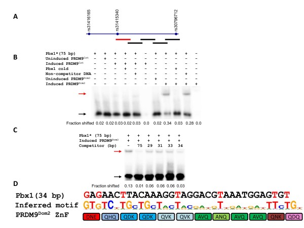
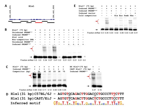
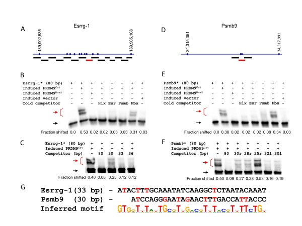
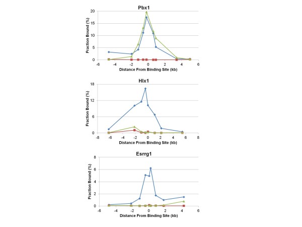
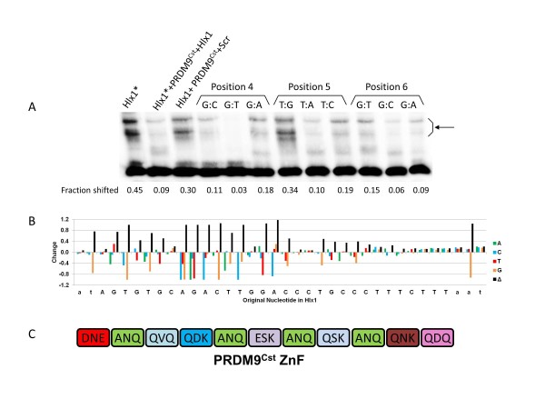
Results: Mouse PRDM9 protein variants bind to hotspot DNA sequences in a manner that is specific for both PRDM9 and DNA haplotypes, and that in vitro binding parallels its in vivo biological activity. Examining four hotspots, three activated by Prdm9Cst and one activated by Prdm9Dom2, we found that all binding sites required the full array of 11 or 12 contiguous fingers, depending on the allele, and that there was little sequence similarity between the binding sites of the three Prdm9Cst activated hotspots. The binding specificity of each position in the Hlx1 binding site, activated by Prdm9Cst, was tested by mutating each nucleotide to its three alternatives. The 31 positions along the binding site varied considerably in the ability of alternative bases to support binding, which also implicates a role for additional binding to the DNA phosphate backbone.

Conclusions: These results, which provide the first detailed mapping of PRDM9 binding to DNA and, to our knowledge, the most detailed analysis yet of DNA binding by a long zinc-finger array, make clear that the binding specificities of PRDM9, and possibly other long-array zinc-finger proteins, are unusually complex.


Figures

Figure 1
Figure 1
Mapping the PRDM9Dom2 binding site of Pbx1. (A) Scheme of Pbx1 hotspot with polymorphisms and amplicons for initial testing of PRDM9Dom2 binding. The red line shows the position of the single binding site detected at this hotspot. (B) Pbx1 specifically binds to PRDM9Dom2 but not PRDM9Cst. The compositions of the binding reactions are shown above each lane. Red arrow, shifted band; black arrow, unbound fragment. (C) Detailed mapping of the Pbx1 binding site. The compositions of the binding reactions are shown above each lane. For the sequences of the competitor oligos used for final mapping, see Additional file 11 (Additional experimental procedures). Red arrow, shifted band; black arrow, unbound fragment. (D) The sequence of the 34 bp oligo representing the minimal binding site is aligned to the inferred PRDM9Dom2 binding motif [6,21] and the zinc-finger array of PRDM9Dom2 (amino acids at positions -1, 3, and 6 relative to the α-helix of each finger are shown). The strong matches of the binding site to the motif are in red.
Figure 2
Figure 2
Defining the PRDM9Cst binding site of Hlx1. (A) Scheme of Hlx1 hotspot with polymorphisms between the C57BL/6J (B6) and the CAST/EiJ (CAST) mouse strains (from Paigen et al. [5]) and amplicons for initial testing of PRDM9Cst binding. The positions of single-nucleotide polymorphisms (SNPs) were taken from the National Center for Biotechnology Information (NCBI) build 37. The red line represents the only fragment showing binding at Hlx1. (B) Hlx1 specifically binds to PRDM9Cst but not PRDM9Dom2. The compositions of the binding reactions are shown above each lane. Red arrow, shifted band; black arrow, unbound fragment. (C) Detailed mapping of the Hlx1 binding site. The compositions of the binding reactions are shown above each lane. For the sequences of the competitor oligos used for final mapping, see Additional file 11 (Additional experimental procedures). (Left panel) Definition of the minimal binding site; (right panel) Strength of binding of PRDM9Cst to B6 and CAST sequences of the Hlx1 binding site. Red arrow, shifted band; black arrow, unbound fragment. (D) Sequences of the Hlx1 minimal binding sites in B6 and CAST strains aligned to the inferred PRDM9Cst binding motif. Sequence differences are underlined. The strong matches of the binding sites to the motif are in red. (E) Hlx1 competes with the other PRDM9Cst-dependent binding sites. The compositions of the binding reactions are shown above each lane. Red arrow, shifted band; black arrow, unbound fragment.
Figure 3
Figure 3
Defining the PRDM9Cst binding sites of Esrrg-1 and Psmb9. (A) Scheme of Esrrg-1 hotspot with polymorphisms between C57BL/6J (B6) and CAST/EiJ (CAST) and amplicons for initial testing of PRDM9Cst binding. The red line represents the only fragment showing binding at this hotspot. (B) Esrrg-1 specifically binds to PRDM9Cst but not PRDM9Dom2 and competes with the other PRDM9Cst-dependent binding sites. The compositions of the binding reactions are shown above each lane. Red arrow, shifted band; black arrow, unbound fragment. (C) Detailed mapping of the Esrrg-1 binding site. The compositions of the binding reactions are shown above each lane. For the sequences of the competitor oligos used for final mapping, see Additional file 11 (Additional experimental procedures). Red arrow, shifted band; black arrow, unbound fragment. (D) Scheme of Psmb9 hotspot with polymorphisms between B6 and CAST, and amplicons for initial testing of PRDM9Cst binding. The approximate position of the Psmb9 binding site was reported previously [19], and therefore only three amplicons surrounding it were tested. Only the middle fragment showed binding to PRDM9Cst. (E) Psmb9 specifically binds to PRDM9Cst but not PRDM9Dom2 and competes with the other PRDM9Cst-dependent binding sites. The compositions of the binding reactions are shown above each lane. Red arrow, shifted band; black arrow, unbound fragment. (F) Detailed mapping of the Psmb9 binding site. The compositions of the binding reactions are shown above each lane. For the sequences of the competitor oligos used for final mapping, see Additional file 11 (Additional experimental procedures). Red arrow, shifted band; black arrow, unbound fragment. (G) Sequences of the Esrrg-1 and Psmb9 minimal binding sites aligned with the inferred PRDM9Cst binding motif. The strong matches of the binding sites to the motif are in red.
Figure 4
Figure 4
H3K4-me3 marks are enriched near the binding sites of the hotspot tested. The peak of H3K4-me3 at hotspots is centered near the PRDM9 binding site. Chromatin was prepared using spermatocytes from mice 12 days post-partum and subjected to chromatin immunoprecipitation (ChIP) with antibody directed to H3K4-me3 or normal rabbit IgG. Quantitative PCR was performed for 8 to 9 amplicons distributed across about 10 kb surrounding the hotspot on immunoprecipitated chromatin and an equal amount of MNase-treated, undiluted input DNA to calculate the fraction of chromatin bound at each amplicon. Blue line, B6 × CAST F1; green line, B6; red line, rabbit IgG (negative control). An identical distribution of H3K4-me3 marks for Psmb9 was shown previously by Grey et al. [19].
Figure 5
Figure 5
Nucleotide substitution analysis of Hlx1-B6 binding to PRDM9Cst. (A) Competition assay for testing the binding of PRDM9Cst to mutated Hlx1 binding sites. Substitutions of nucleotides 4 to 6; labeled oligos are indicated by asterisk. The compositions of reaction mixtures in the first three lanes are shown above each lane. Lanes 4 to 12 show the competition test with mutated oligos. The position and the nature of each mutation in the oligo are shown. The composition of the reaction mixtures in lanes 4 to 12 is mutated oligo + PRDM9Cst + oligo* (see Materials and methods). The fraction of the shifted band is indicated below each lane. Red arrow, shifted band; black arrow, unbound fragment. (B) Graphic representation of the binding changes at each position produced by the nucleotide substitution analysis. (C) ZF domain of PRDM9Cst. The letters in the boxes show the amino acids at positions -1, 3, and 6 relative to the α-helix of each finger.

Comment in

  • The complex binding of PRDM9.
    Ségurel L. Ségurel L. Genome Biol. 2013 Apr 24;14(4):112. doi: 10.1186/gb-2013-14-4-112. Genome Biol. 2013. PMID: 23651476 Free PMC article.

References

    1. Petes TD. Meiotic recombination hot spots and cold spots. Nat Rev Genet. 2001;14:360–369. doi: 10.1038/35072078. - DOI - PubMed
    1. Drouaud J, Camilleri C, Bourguignon PY, Canaguier A, Berard A, Vezon D, Giancola S, Brunel D, Colot V, Prum B, Quesneville H, Mézard C. Variation in crossing-over rates across chromosome 4 of Arabidopsis thaliana reveals the presence of meiotic recombination "hot spots". Genome Res. 2006;14:106–114. - PMC - PubMed
    1. Jeffreys AJ, Kauppi L, Neumann R. Intensely punctate meiotic recombination in the class II region of the major histocompatibility complex. Nat Genet. 2001;14:217–222. doi: 10.1038/ng1001-217. - DOI - PubMed
    1. de Massy B. Distribution of meiotic recombination sites. Trends Genet. 2003;14:514–522. doi: 10.1016/S0168-9525(03)00201-4. - DOI - PubMed
    1. Paigen K, Szatkiewicz JP, Sawyer K, Leahy N, Parvanov ED, Ng SH, Graber JH, Broman KW, Petkov PM. The recombinational anatomy of a mouse chromosome. PLoS Genet. 2008;14:e1000119. doi: 10.1371/journal.pgen.1000119. - DOI - PMC - PubMed

Publication types