. 2013 May 1;27(9):1003-15.
doi: 10.1101/gad.211326.112. Epub 2013 Apr 25.

The retinoblastoma protein induces apoptosis directly at the mitochondria

Affiliations

The retinoblastoma protein induces apoptosis directly at the mitochondria

Keren I Hilgendorf et al. Genes Dev. .

Abstract

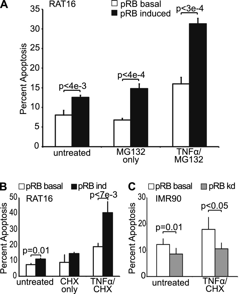
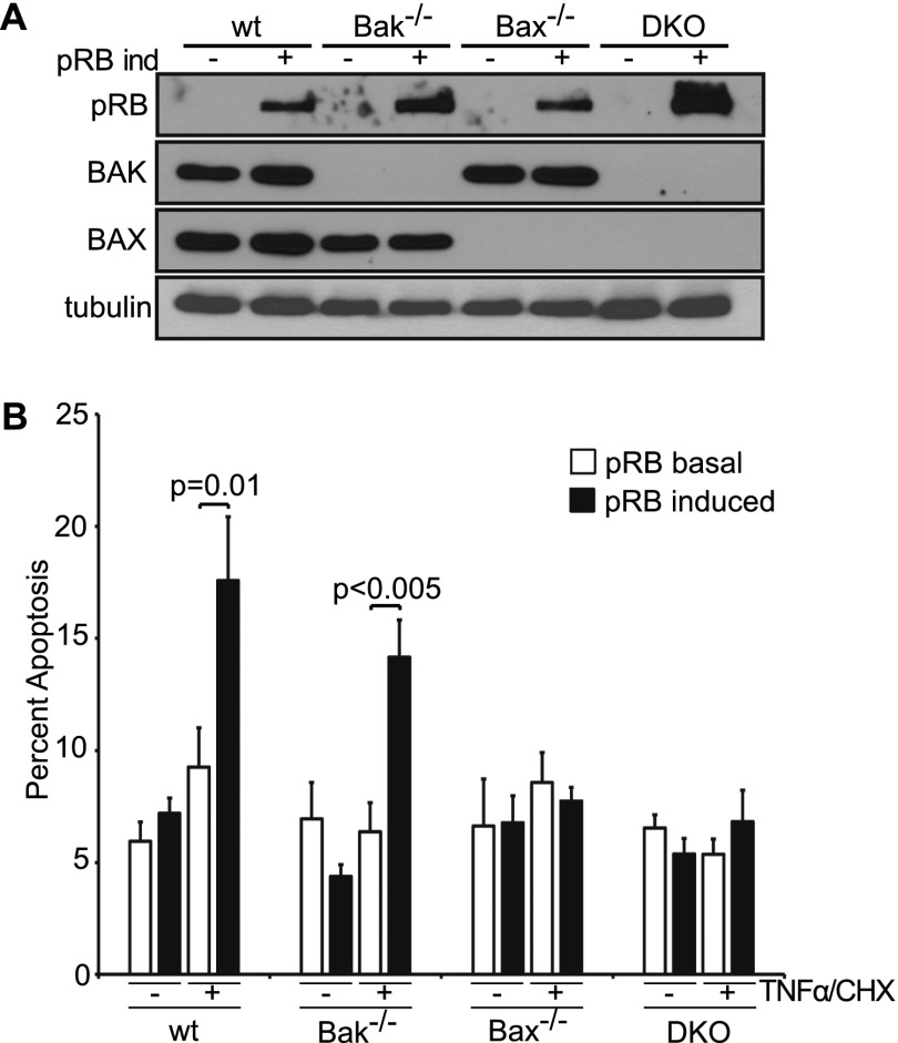
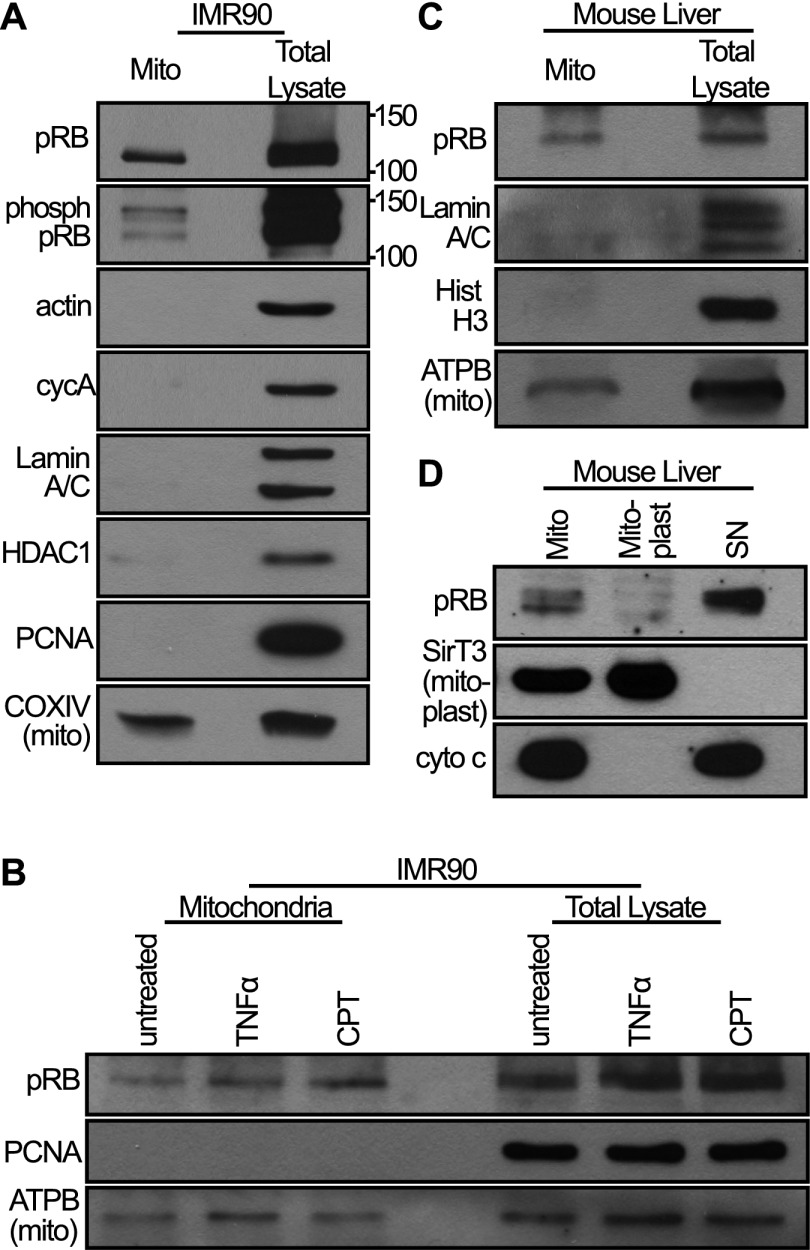
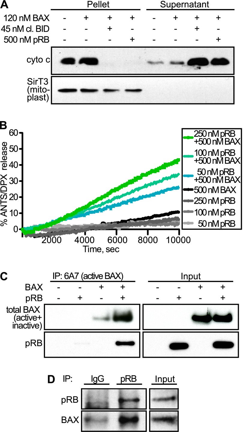
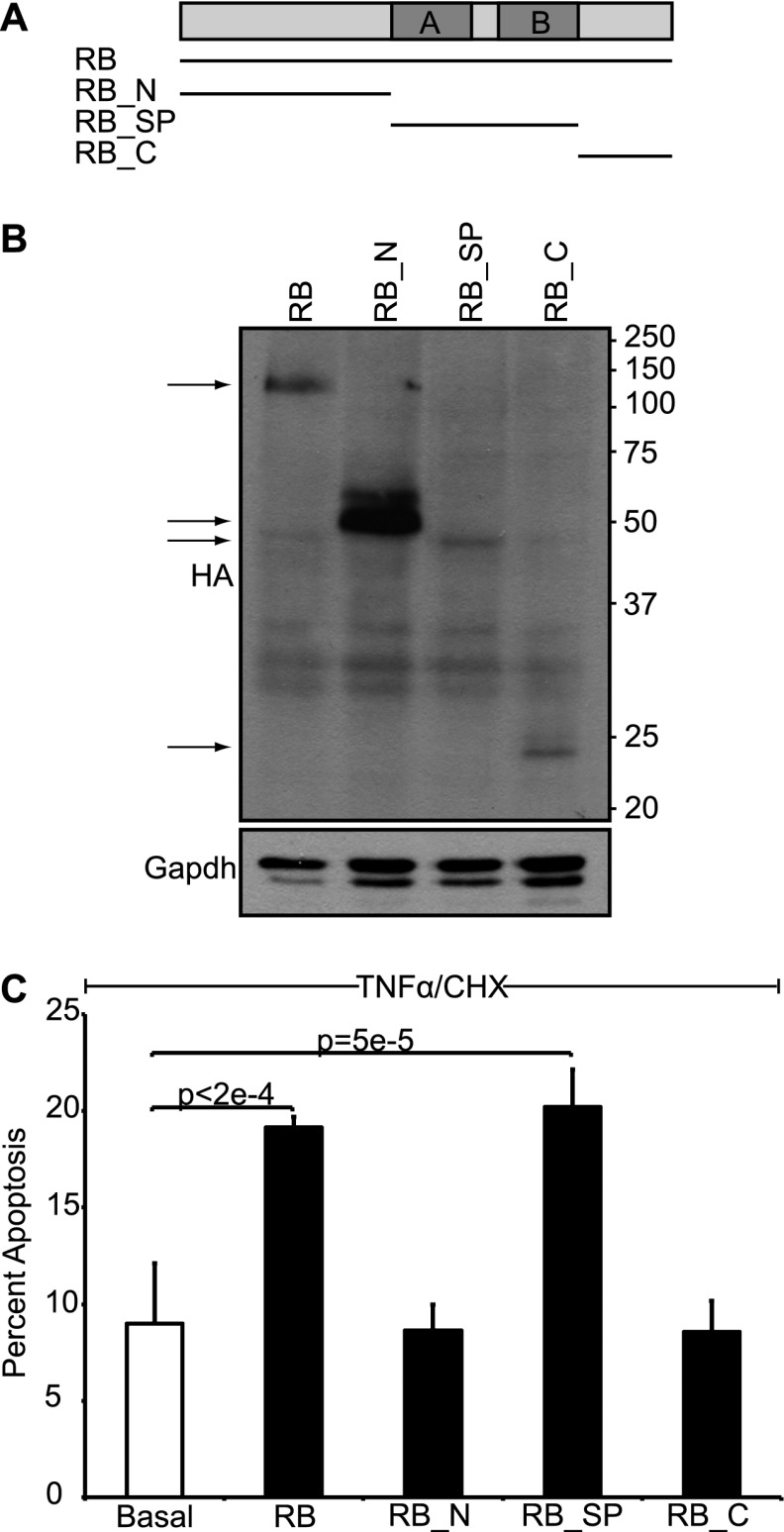
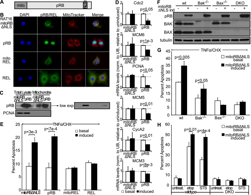
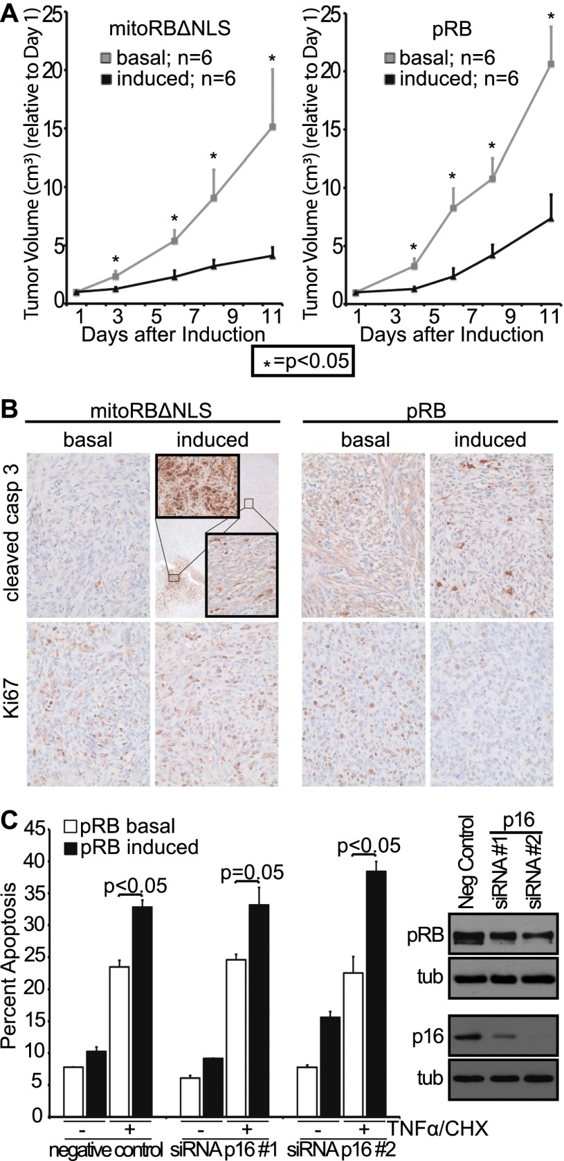
The retinoblastoma protein gene RB-1 is mutated in one-third of human tumors. Its protein product, pRB (retinoblastoma protein), functions as a transcriptional coregulator in many fundamental cellular processes. Here, we report a nonnuclear role for pRB in apoptosis induction via pRB's direct participation in mitochondrial apoptosis. We uncovered this activity by finding that pRB potentiated TNFα-induced apoptosis even when translation was blocked. This proapoptotic function was highly BAX-dependent, suggesting a role in mitochondrial apoptosis, and accordingly, a fraction of endogenous pRB constitutively associated with mitochondria. Remarkably, we found that recombinant pRB was sufficient to trigger the BAX-dependent permeabilization of mitochondria or liposomes in vitro. Moreover, pRB interacted with BAX in vivo and could directly bind and conformationally activate BAX in vitro. Finally, by targeting pRB specifically to mitochondria, we generated a mutant that lacked pRB's classic nuclear roles. This mito-tagged pRB retained the ability to promote apoptosis in response to TNFα and also additional apoptotic stimuli. Most importantly, induced expression of mito-tagged pRB in Rb(-/-);p53(-/-) tumors was sufficient to block further tumor development. Together, these data establish a nontranscriptional role for pRB in direct activation of BAX and mitochondrial apoptosis in response to diverse stimuli, which is profoundly tumor-suppressive.

Keywords: MOMP; apoptosis; cancer; pRB; retinoblastoma protein.


Figures

Figure 1.
Figure 1.
pRB promotes TNFα-induced apoptosis in a transcription-independent manner. (A) RAT16 cells with and without induced expression of pRB for 24 h were treated with TNFα and MG132 for 48 h and analyzed for apoptosis by AnnexinV staining. Induced expression of pRB resulted in increased levels of apoptosis without TNFα treatment and greatly enhanced TNFα/MG132-induced apoptosis. (B) Induced expression of pRB for 24 h in RAT16 cells increased apoptosis resulting from 24 h of TNFα and CHX treatment. (C) Stable knockdown of pRB in IMR90 cells decreased apoptosis induced by 24 h of treatment with TNFα and CHX. (A–C) Graph bars represent the average of at least three independent experiments (±SD).
Figure 2.
Figure 2.
pRB promotes mitochondrial apoptosis in a BAX-dependent manner. (A) pRB expression was induced for 24 h in stable pools of wild-type, Bak−/−, Bax−/−, and Bax−/−;Bak−/− immortalized MEFs by doxycycline addition and confirmed by Western blotting using antibodies against pRB, BAK, BAX, and tubulin. (B) Wild-type, Bak−/−, Bax−/−, and Bax−/−;Bak−/− immortalized MEFs with or without 24 h of pRB expression were left untreated or treated with TNFα and CHX for 10 h and analyzed for apoptosis by AnnexinV staining. Induction of pRB in wild-type and Bak−/−, but not Bax−/− or Bax−/−;Bak−/−, MEFs sensitized to TNFα/CHX-induced apoptosis. Each MEF variant was independently generated twice. Graph bars represent the average of three representative, independent experiments (±SD).
Figure 3.
Figure 3.
A fraction of pRB constitutively localizes to mitochondria. (A) IMR90 cell mitochondria were fractionated, and equal amounts (in micrograms) of mitochondrial fraction (mito) versus total lysate were analyzed by Western blotting using an antibody against pRB and a cocktail of antibodies against phosphorylated pRB. A fraction of pRB, including phospho-pRB, localizes to mitochondria. The purity of the mitochondrial fraction was verified using control nuclear and cytoplasmic markers. (B) Mitochondria of IMR90 cells treated with camptothecin (CPT; 5 h) or TNFα (24 h) were isolated and analyzed by Western blotting. The levels of mitochondrial pRB are unaffected by treatment with these drugs. (C) Mitochondria were isolated from mouse livers and analyzed by Western blotting. A fraction of pRB is present at mouse liver mitochondria. (D) Mouse liver mitochondria were subfractionated into mitoplast and nonmitoplast (SN). Mitochondrial pRB localizes outside the mitoplast. (A–D) Data are representative of at least three independent experiments.
Figure 4.
Figure 4.
pRB induces MOMP by directly activating BAX. (A) Mouse liver mitochondria were isolated; supplemented with recombinant, monomeric BAX; and incubated with and without recombinant cleaved BID (positive control) or baculovirus-expressed, recombinant human pRB. Cytochrome c release was assessed following incubation and centrifugation by Western blotting of pellet versus supernatant. Addition of either pRB or cleaved BID was sufficient to release cytochrome c into the supernatant. (B) ANTS/DPX-loaded liposomes were incubated with 50, 100, and 250 nM recombinant pRB in the presence or absence of recombinant, monomeric BAX. ANTS/DPX release was assessed over time. pRB yielded dose-dependent liposome permeabilization in a BAX-dependent manner. (C) In vitro binding assay using recombinant pRB and recombinant, monomeric BAX. Activated BAX was immunoprecipitated using an active conformation-specific BAX antibody (6A7), and binding was assessed by Western blotting using an antibody against total BAX (inactive + active) and pRB. When coincubated with inactive BAX, pRB bound to (bottom panels) and stimulated formation of (top panels) conformationally active BAX. (D) Coimmunoprecipitation experiment using IMR90 cells treated with TNFα/CHX for 3 h. Endogenous pRB was immunoprecipitated from cell extracts, and endogenous BAX association was assessed by Western blotting. pRB and BAX form an endogenous complex in vivo in TNFα/CHX-treated cells. (A–D) Data are representative of at least three independent experiments.
Figure 5.
Figure 5.
The small pocket of pRB is sufficient to induce transcription-independent apoptosis. (A) Schematic of pRB domains: the N terminus (RB_N; residues 1–372), the small pocket domain (RB_SP; residues 373–766), and the C terminus (RB_C; residues 767–928). (B) Stable variants of RAT16 cells allowing for inducible expression of HA-tagged full-length pRB, RB_N, RB_SP, and RB_C by doxycycline withdrawal were generated, and expression was verified after 24 h by Western blotting using an HA-antibody. (C) RAT16 cells with and without expression of pRB, RB_N, RB_SP, and RB_C for 24 h were treated with TNFα and CHX for 24 h, and levels of apoptosis were assessed by AnnexinV staining. Induced expression of full-length pRB and RB_SP, but not RB_N or RB_C, sensitized to TNFα/CHX-induced apoptosis. (B,C) Each RAT16 variant was independently generated three times. Graph bars represent the average of three representative, independent experiments (±SD).
Figure 6.
Figure 6.
Mitochondria targeted pRB is deficient for pRB's nuclear function but induces apoptosis in response to various apoptotic stimuli. (A) Schematic of mitoRBΔNLS construct. pRB was targeted to mitochondria by fusion to the mitochondrial leader peptide of ornithine transcarbamylase and mutation of the NLS. (B) Stable variants of RAT16 cells allowing for inducible expression of mitoRBΔNLS, wild-type pRB, mitoREL, and wild-type REL were generated, and cellular localization following 24 h of induction was assessed by immunofluorescence using antibodies against pRB and REL. Mitochondria were visualized using MitoTracker. mitoRBΔNLS, and mitoREL localized to mitochondria. (C) Western blotting showing induced expression levels of mitoRBΔNLS and wild-type pRB at mitochondria and total lysate. mitoRBΔNLS expressed at lower levels than wild-type pRB even when considering the mitochondrial fraction. (D) pRB, but not mitoRBΔNLS, repressed E2F target genes cdc2, mcm3, mcm5, mcm6, PCNA, and cycA2 as measured by RT-qPCR and normalized to ubiquitin. Average of two independent experiments (±SD). (E) Induced expression of mitoRBΔNLS and wild-type pRB, but not mitoREL or REL, sensitized to apoptosis induced by 24 h of treatment with TNFα and CHX. (B–E) Each RAT16 variant was independently generated three times. Graph bars represent the average of three representative, independent experiments (±SD). (F) mitoRBΔNLS expression was induced by doxycycline addition for 24 h in stable variants of wild-type, Bak−/−, Bax−/−, and Bax−/−;Bak−/− immortalized MEFs and confirmed by Western blotting using antibodies against pRB, BAK, BAX, and tubulin. (G) Induction of mitoRBΔNLS in wild-type and Bak−/−, but not Bax−/− or Bax−/−;Bak−/−, MEFs sensitized to 10 h of treatment with TNFα and CHX. (H) Wild-type and Bax−/−;Bak−/− MEFs with and without induced expression of mitoRBΔNLS were treated with STS (1 μM) for 6 h or etoposide (25 μM) for 12 h. Induction of mitoRBΔNLS in wild-type, but not Bax−/−;Bak−/−, MEFs enhanced apoptosis in response to STS and etoposide. (F–H) Each MEF variant was independently generated twice. Graph bars represent the average of three representative, independent experiments (±SD).
Figure 7.
Figure 7.
Mitochondria targeted pRB induces apoptosis in vivo and is tumor-suppressive. (A) Rb−/−;p53−/− osteosarcoma cell variants allowing for doxycycline-inducible expression of mitoRBΔNLS or wild-type pRB were injected into the flanks of nude mice (two injection sites per mouse; 12 injections per cell line variant). Once a tumor volume of ∼0.05 cm3 was reached, mitoRBΔNLS or wild-type pRB expression was induced in half the mice, and tumor volume was monitored for 11 d (n = 6 per condition, ±SEM). Induced expression of mitoRBΔNLS or wild-type pRB suppressed growth of xenografted tumors. Tumor volume normalized to day 1. (B, top) Induced expression of mitoRBΔNLS or wild-type pRB resulted in increased levels of apoptosis as measured by immunohistochemistry using a cleaved caspase 3 antibody. (Inset) Tumors expressing mitoRBΔNLS also contained small areas with very high levels of apoptosis. (Bottom) Induced expression of wild-type pRB, but not mitoRBΔNLS, decreased proliferation as measured by Ki67 staining. Images representative of six respective tumor sections. (C) Murine p16 was knocked down in immortalized wild-type MEF variants that allow for doxycycline-inducible expression of pRB. Phosphorylation status of pRB (as judged by mobility) and knockdown of p16 using two distinct siRNAs were confirmed by Western blotting. Induced expression of pRB enhances TNFα/CHX-induced apoptosis in the presence and absence of p16, and this activity is not inactivated by pRB phosphorylation. Bars indicate the average of two independent experiments (±SD).

Comment in

  • RB goes mitochondrial.
    Attardi LD, Sage J. Attardi LD, et al. Genes Dev. 2013 May 1;27(9):975-9. doi: 10.1101/gad.219451.113. Genes Dev. 2013. PMID: 23651852 Free PMC article.

References

    1. Araki K, Ahmad SM, Li G, Bray DA Jr, Saito K, Wang D, Wirtz U, Sreedharan S, O'Malley BW Jr, Li D 2008. Retinoblastoma RB94 enhances radiation treatment of head and neck squamous cell carcinoma. Clin Cancer Res 14: 3514–3519 - PubMed
    1. Blanchet E, Annicotte J-Sb, Lagarrigue S, Aguilar V, Clapé C, Chavey C, Fritz V, Casas Fo, Apparailly F, Auwerx J, et al. 2011. E2F transcription factor-1 regulates oxidative metabolism. Nat Cell Biol 13: 1146–1152 - PMC - PubMed
    1. Bosco EE, Mayhew CN, Hennigan RF, Sage J, Jacks T, Knudsen ES 2004. RB signaling prevents replication-dependent DNA double-strand breaks following genotoxic insult. Nucleic Acids Res 32: 25–34 - PMC - PubMed
    1. Brunelle J, Letai A 2009. Control of mitochondrial apoptosis by the Bcl-2 family. J Cell Sci 122: 437–478 - PMC - PubMed
    1. Burkhart DL, Sage J 2008. Cellular mechanisms of tumour suppression by the retinoblastoma gene. Nat Rev Cancer 8: 671–682 - PMC - PubMed

Publication types