. 2013 Apr 25;4(4):e608.
doi: 10.1038/cddis.2013.129.

NMDA receptor subunit composition determines beta-amyloid-induced neurodegeneration and synaptic loss

Affiliations

NMDA receptor subunit composition determines beta-amyloid-induced neurodegeneration and synaptic loss

C Tackenberg et al. Cell Death Dis. .

Abstract

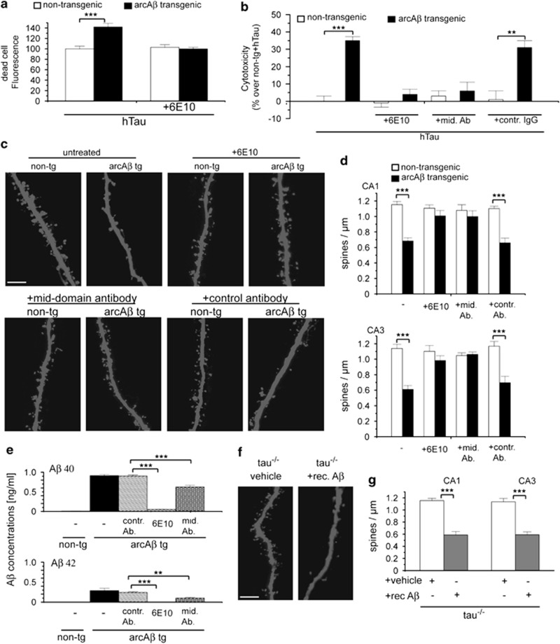
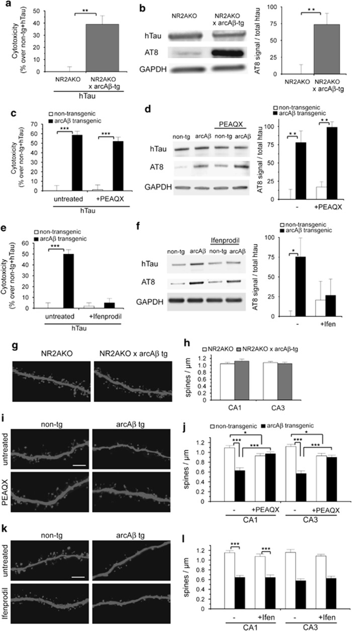
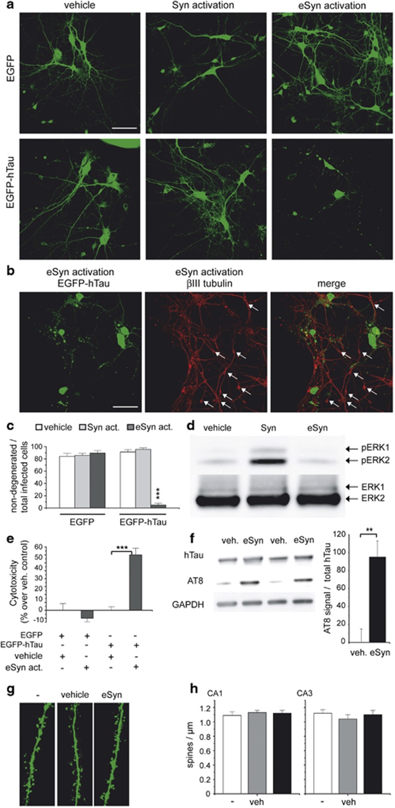
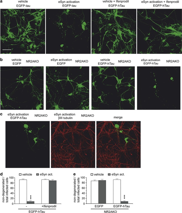
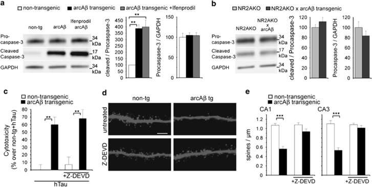
Aggregates of amyloid-beta (Aβ) and tau are hallmarks of Alzheimer's disease (AD) leading to neurodegeneration and synaptic loss. While increasing evidence suggests that inhibition of N-methyl-D-aspartate receptors (NMDARs) may mitigate certain aspects of AD neuropathology, the precise role of different NMDAR subtypes for Aβ- and tau-mediated toxicity remains to be elucidated. Using mouse organotypic hippocampal slice cultures from arcAβ transgenic mice combined with Sindbis virus-mediated expression of human wild-type tau protein (hTau), we show that Aβ caused dendritic spine loss independently of tau. However, the presence of hTau was required for Aβ-induced cell death accompanied by increased hTau phosphorylation. Inhibition of NR2B-containing NMDARs abolished Aβ-induced hTau phosphorylation and toxicity by preventing GSK-3β activation but did not affect dendritic spine loss. Inversely, NR2A-containing NMDAR inhibition as well as NR2A-subunit knockout diminished dendritic spine loss but not the Aβ effect on hTau. Activation of extrasynaptic NMDARs in primary neurons caused degeneration of hTau-expressing neurons, which could be prevented by NR2B-NMDAR inhibition but not by NR2A knockout. Furthermore, caspase-3 activity was increased in arcAβ transgenic cultures. Activity was reduced by NR2A knockout but not by NR2B inhibition. Accordingly, caspase-3 inhibition abolished spine loss but not hTau-dependent toxicity in arcAβ transgenic slice cultures. Our data show that Aβ induces dendritic spine loss via a pathway involving NR2A-containing NMDARs and active caspase-3 whereas activation of eSyn NR2B-containing NMDARs is required for hTau-dependent neurodegeneration, independent of caspase-3.


Figures

Figure 1
Figure 1
Aβ induces hTau-dependent neurotoxicity and tau-independent spine loss. (a) Quantification of dead cell fluorescence intensities of hTau-expressing hippocampal slice cultures from arcAβ tg and non-tg mice treated with Aβ antibody 6E10 determined by Live/Dead cell viability/cytotoxicity assay. (b) Cytotoxicity of hTau in arcAβ tg and non-tg control slice cultures treated with 1 μg/ml of Aβ antibody 6E10, a mid-domain Aβ antibody or control antibody measured by Cytotox-Glo assay. (c) Confocal images of apical dendritic segments from CA1 neurons of arcAβ tg and non-tg slices in the presence or absence of Aβ antibody 6E10, mid-domain Aβ antibody or control antibody. (d) Quantification of spine density in cultures from arcAβ tg and non-tg mice. (e) Quantitative bar graphs representing mean values of the amount of Aβ40 and Aβ42 peptides in medium of arcAβ hippocampal slice cultures after treatment with respective Aβ antibodies as determined by ELISA. (f) Representative images of apical dendritic segments from CA1 neurons from tau−/− mice treated with 1 μM recombinant Aβ42. (g) Quantification of spine density in tau−/− cultures treated with recombinant Aβ42. rec. Aβ, recombinant Aβ42; mid. Ab, mid-domain Aβ antibody; contr. Ab, control antibody; mean±S.E.M.; **P<0.01 and ***P<0.001; Mann–Whitney-U-test; n=9–13, n=4 (e) scale bar: 5 μm
Figure 2
Figure 2
NR2B-containing NMDAR inhibition prevents Aβ-induced hTau-dependent toxicity whereas NR2A knockout or inhibition abolishes dendritic spine loss. (a) Cytotoxicity of hTau in NR2AKO × arcAβ tg and NR2AKO cultures measured by Cytotox-Glo assay. (b) Western blot showing AT8 phosphorylation of hTau in NR2AKO × arcAβ tg and NR2AKO cultures. (c) Cytotoxicity of hTau in arcAβ tg and non-tg control cultures treated with 50 nM PEAQX. (d) Western blot showing expression of hTau and phosphorylation at AT8 epitope after PEAQX treatment. (e) Cytotoxicity of hTau in arcAβ tg and non-tg control cultures treated with 3 μM Ifenprodil. (f) Western blot showing phosphorylation of hTau at AT8 epitope after Ifenprodil treatment. (g) Representative confocal images of apical dendritic segments from NR2AKO and NR2AKO × arcAβ tg cultures. (h) Spine density in NR2AKO and NR2AKO × arcAβ tg cultures. (i) Apical dendritic segments from CA1 neurons of arcAβ tg and non-tg slices in the presence or absence of 50 nM PEAQX. (j) Quantification of spine density after treatment with PEAQX. (k) Apical dendritic segments from CA1 neurons of arcAβ tg and non-tg slices in the presence or absence of 3 μM Ifenprodil. (l) Quantification of spine density after treatment with Ifenprodil. Ifen, Ifenprodil; NR2AKO, NR2A-containing NMDAR knockout; values are shown as mean±S.E.M. with *P<0.05, **P<0.01 and ***P<0.001; Mann–Whitney-U-test; n=6–8 (a, c, e), n=5 (b), n=4 (d), n=6 (f), n=10–13 (h, j, l); scale bar: 5 μm
Figure 3
Figure 3
Extrasynaptic NMDAR activation induces hTau-dependent toxicity in primary neuronal cultures and hippocampal slice cultures. (a) Representative confocal images of primary neurons expressing EGFP or EGFP-hTau after synaptic or extrasynaptic activation. (b) Confocal images of EGFP-hTau-expressing neurons after extrasynaptic activation and immunostaining against βIII tubulin. Arrows mark non-infected neurons. (c) Quantification of hTau-dependent toxicity. Shown is the ratio of non-degenerated infected primary neurons (neurons without fragmented or beaded neurites or ballooned morphology) to total infected neurons. (d) Representative western blot with phospho-ERK 1/2 (pERK) and ERK 1/2 antibodies of lysates from primary neurons after synaptic or extrasynaptic activation. (e) Cytotoxicity of hTau in wt hippocampal slice cultures after extrasynaptic activation measured by Cytotox-Glo assay. (f) Western blot showing AT8 phosphorylation of hTau after extrasynaptic activation in slice cultures. (g) Representative dendritic segments from CA1 neurons of wt slice cultures after extrasynaptic activation. (h) Quantification of spine density in wt slices analyzed 1 day after extrasynaptic activation. eSyn, extrasynaptic; Syn, synaptic activation; veh, vehicle; values are shown as mean±S.E.M. with **P<0.01 and ***P<0.001; Mann–Whitney-U-test; n=10–13 (c), n=3 (d); n=8 (e), n=4 (f); n=10 (h) scale bars: 50 μm (a, b), 5 μm (g)
Figure 4
Figure 4
NR2B-containing NMDAR inhibition but not NR2A-containing NMDAR knockout abolishes hTau-dependent toxicity after activation of extrasynaptic NMDARs. (a) Representative confocal images of primary neurons expressing EGFP-hTau after extrasynaptic activation in the presence and absence of 3 μM Ifenprodil. (b) Representative confocal images of primary neurons from NR2AKO mice expressing EGFP-hTau after extrasynaptic activation. (c) Confocal images of EGFP-hTau-expressing primary neurons from NR2AKO mice after extrasynaptic activation and immunostaining against βIII tubulin. (d) Quantification of hTau-dependent toxicity in the presence and absence of Ifenprodil. Shown is the ratio of non-degenerated infected neurons (neurons without fragmented or beaded neurites or ballooned morphology) to total infected neurons. (e) Quantification of hTau-dependent toxicity in primary neurons of NR2AKO mice. eSyn act, activated eSyn NMDARs; values are shown as mean±S.E.M. with ***P<0.001; Mann–Whitney-U-test; n=10–14; scale bar: 50 μm
Figure 5
Figure 5
Aβ-induced caspase-3 activation causes dendritic spine loss but not hTau-dependent toxicity. (a) Western blot with antibodies against procaspase-3 and cleaved (activated) caspase-3 in slice cultures from arcAβ tg mice treated with 3 μM Ifenprodil. (b) Western blot with antibodies against procaspase-3 and cleaved caspase-3 in cultured slices from NR2AKO and NR2AKO × arcAβ tg mice. (c) Cytotoxicity of hTau in arcAβ tg and non-tg control slice cultures treated with 10 μM Z-DEVD-FMK measured by Cytotox-Glo assay. (d) Representative confocal images of apical dendritic segments from CA1 neurons of arcAβ tg and non-tg slices treated with 10 μM Z-DEVD-FMK. (e) Quantification of spine density after treatment with Z-DEVD-FMK. Z-DEVD, Z-DEVD-FMK; values are shown as mean±S.E.M. with **P<0.01 and ***P<0.001; Mann–Whitney-U-test; n=3–4 (a, b), n=7–11 (c, e); scale bar: 5 μm

Similar articles

Cited by

References

    1. Rapoport M, Dawson HN, Binder LI, Vitek MP, Ferreira A. Tau is essential to beta -amyloid-induced neurotoxicity. Proc Natl Acad Sci USA. 2002;99:6364–6369. - PMC - PubMed
    1. Tackenberg C, Brandt R. Divergent pathways mediate spine alterations and cell death induced by amyloid-beta, wild-type tau, and R406W tau. J Neurosci. 2009;29:14439–14450. - PMC - PubMed
    1. Roberson E, Scearce-Levie K, Palop JJ, Yan F, Cheng IH, Wu T, et al. Reducing endogenous tau ameliorates amyloid beta-induced deficits in an Alzheimer's disease mouse model. Science. 2007;31:750–754. - PubMed
    1. Ittner LM, Ke YD, Delerue F, Bi M, Gladbach A, van Eersel J, et al. Dendritic function of tau mediates amyloid-beta toxicity in Alzheimer's disease mouse models. Cell. 2010;142:387–397. - PubMed
    1. Nussbaum JM, Schilling S, Cynis H, Silva A, Swanson E, Wangsanut T, et al. Prion-like behaviour and tau-dependent cytotoxicity of pyroglutamylated amyloid-ß. Nature. 2012;485:651–655. - PMC - PubMed

Publication types

MeSH terms