

Assessment of disease activity in muscular dystrophies by noninvasive imaging
- PMID: 23619364
- PMCID: PMC3638910
- DOI: 10.1172/JCI68458
Assessment of disease activity in muscular dystrophies by noninvasive imaging
Abstract
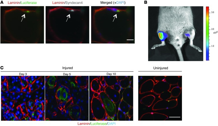
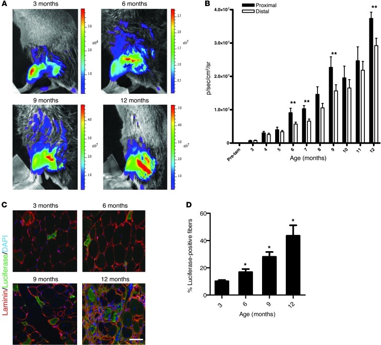
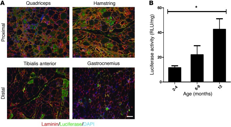
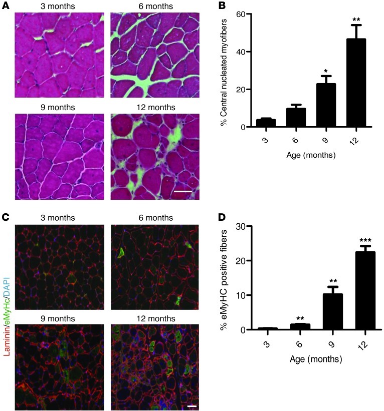
Muscular dystrophies are a class of disorders that cause progressive muscle wasting. A major hurdle for discovering treatments for the muscular dystrophies is a lack of reliable assays to monitor disease progression in animal models. We have developed a novel mouse model to assess disease activity noninvasively in mice with muscular dystrophies. These mice express an inducible luciferase reporter gene in muscle stem cells. In dystrophic mice, muscle stem cells activate and proliferate in response to muscle degeneration, resulting in an increase in the level of luciferase expression, which can be monitored by noninvasive, bioluminescence imaging. We applied this noninvasive imaging to assess disease activity in a mouse model of the human disease limb girdle muscular dystrophy 2B (LGMD2B), caused by a mutation in the dysferlin gene. We monitored the natural history and disease progression in these dysferlin-deficient mice up to 18 months of age and were able to detect disease activity prior to the appearance of any overt disease manifestation by histopathological analyses. Disease activity was reflected by changes in luciferase activity over time, and disease burden was reflected by cumulative luciferase activity, which paralleled disease progression as determined by histopathological analysis. The ability to monitor disease activity noninvasively in mouse models of muscular dystrophy will be invaluable for the assessment of disease progression and the effectiveness of therapeutic interventions.
Figures

Comment in
-
Illuminating regeneration: noninvasive imaging of disease progression in muscular dystrophy.J Clin Invest. 2013 May;123(5):1931-4. doi: 10.1172/JCI69568. Epub 2013 Apr 24. J Clin Invest. 2013. PMID: 23619358 Free PMC article.
Similar articles
-
Illuminating regeneration: noninvasive imaging of disease progression in muscular dystrophy.J Clin Invest. 2013 May;123(5):1931-4. doi: 10.1172/JCI69568. Epub 2013 Apr 24. J Clin Invest. 2013. PMID: 23619358 Free PMC article.
-
Genetic manipulation of dysferlin expression in skeletal muscle: novel insights into muscular dystrophy.Am J Pathol. 2009 Nov;175(5):1817-23. doi: 10.2353/ajpath.2009.090107. Epub 2009 Oct 15. Am J Pathol. 2009. PMID: 19834057 Free PMC article.
-
Dysferlin and the plasma membrane repair in muscular dystrophy.Trends Cell Biol. 2004 Apr;14(4):206-13. doi: 10.1016/j.tcb.2004.03.001. Trends Cell Biol. 2004. PMID: 15066638 Review.
-
Defective membrane repair in dysferlin-deficient muscular dystrophy.Nature. 2003 May 8;423(6936):168-72. doi: 10.1038/nature01573. Nature. 2003. PMID: 12736685
-
Muscular dystrophy in dysferlin-deficient mouse models.Neuromuscul Disord. 2013 May;23(5):377-87. doi: 10.1016/j.nmd.2013.02.004. Epub 2013 Mar 7. Neuromuscul Disord. 2013. PMID: 23473732 Review.
Cited by
-
Plasmid-Mediated Gene Therapy in Mouse Models of Limb Girdle Muscular Dystrophy.Mol Ther Methods Clin Dev. 2019 Oct 14;15:294-304. doi: 10.1016/j.omtm.2019.10.002. eCollection 2019 Dec 13. Mol Ther Methods Clin Dev. 2019. PMID: 31890729 Free PMC article.
-
zebraflash transgenic lines for in vivo bioluminescence imaging of stem cells and regeneration in adult zebrafish.Development. 2013 Dec;140(24):4988-97. doi: 10.1242/dev.102053. Epub 2013 Nov 6. Development. 2013. PMID: 24198277 Free PMC article.
-
Monitoring disease activity noninvasively in the mdx model of Duchenne muscular dystrophy.Proc Natl Acad Sci U S A. 2018 Jul 24;115(30):7741-7746. doi: 10.1073/pnas.1802425115. Epub 2018 Jul 9. Proc Natl Acad Sci U S A. 2018. PMID: 29987034 Free PMC article.
-
Seeing stem cells at work in vivo.Stem Cell Rev Rep. 2014 Feb;10(1):127-44. doi: 10.1007/s12015-013-9468-x. Stem Cell Rev Rep. 2014. PMID: 23975604 Free PMC article. Review.
-
The protein tyrosine phosphatase 1B inhibitor MSI-1436 stimulates regeneration of heart and multiple other tissues.NPJ Regen Med. 2017 Mar 3;2:4. doi: 10.1038/s41536-017-0008-1. eCollection 2017. NPJ Regen Med. 2017. PMID: 29302341 Free PMC article.
References
Publication types
MeSH terms
Substances
Grants and funding
LinkOut - more resources
Full Text Sources
Other Literature Sources
Medical
Molecular Biology Databases
