. 2013 May;123(5):2143-54.
doi: 10.1172/JCI64365. Epub 2013 Apr 24.

MicroRNA-146a is a therapeutic target and biomarker for peripartum cardiomyopathy

Affiliations

MicroRNA-146a is a therapeutic target and biomarker for peripartum cardiomyopathy

Julie Halkein et al. J Clin Invest. 2013 May.

Abstract

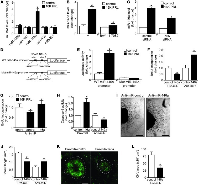
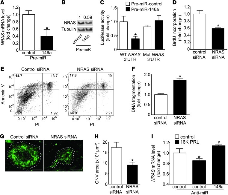
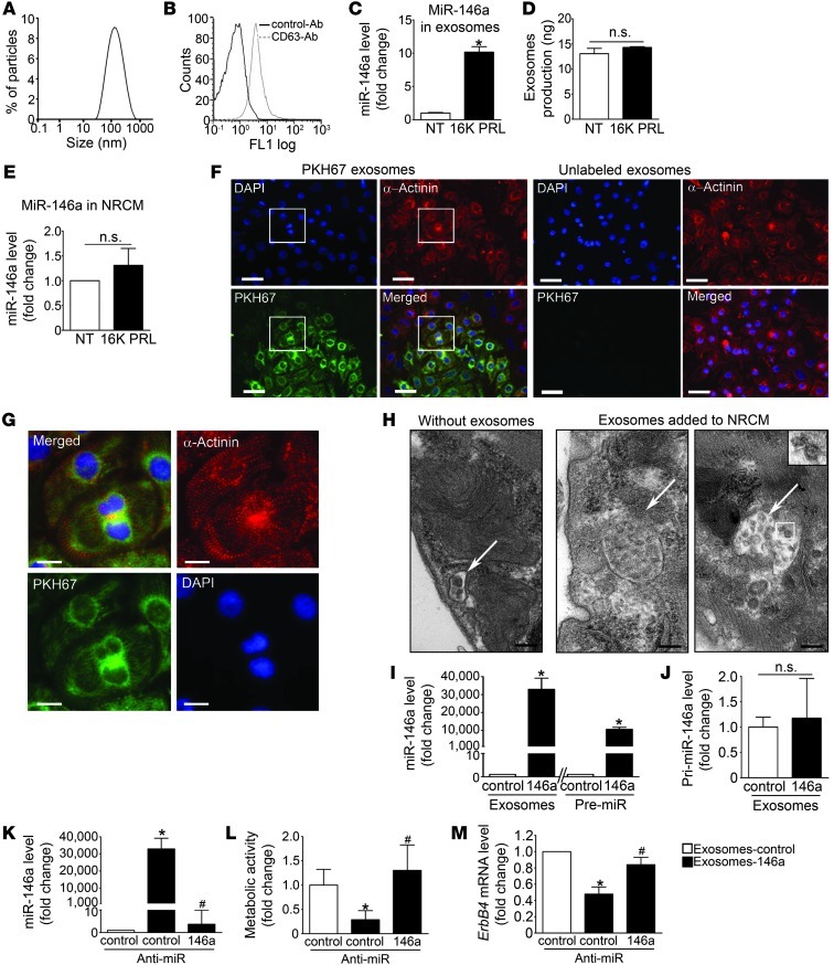
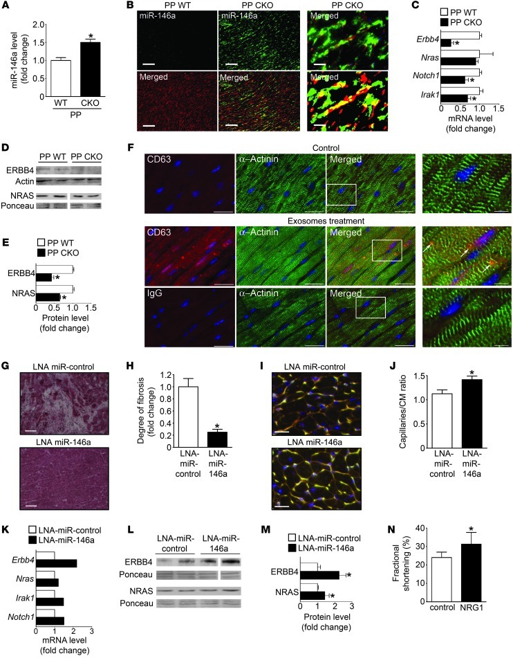
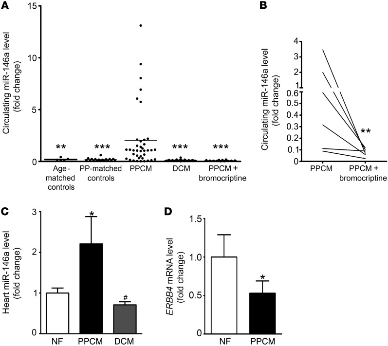
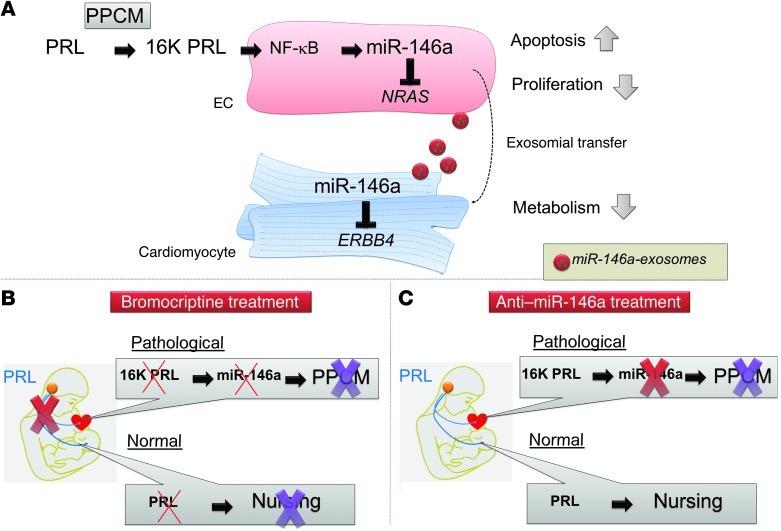
Peripartum cardiomyopathy (PPCM) is a life-threatening pregnancy-associated cardiomyopathy in previously healthy women. Although PPCM is driven in part by the 16-kDa N-terminal prolactin fragment (16K PRL), the underlying molecular mechanisms are poorly understood. We found that 16K PRL induced microRNA-146a (miR-146a) expression in ECs, which attenuated angiogenesis through downregulation of NRAS. 16K PRL stimulated the release of miR-146a-loaded exosomes from ECs. The exosomes were absorbed by cardiomyocytes, increasing miR-146a levels, which resulted in a subsequent decrease in metabolic activity and decreased expression of Erbb4, Notch1, and Irak1. Mice with cardiomyocyte-restricted Stat3 knockout (CKO mice) exhibited a PPCM-like phenotype and displayed increased cardiac miR-146a expression with coincident downregulation of Erbb4, Nras, Notch1, and Irak1. Blocking miR-146a with locked nucleic acids or antago-miRs attenuated PPCM in CKO mice without interrupting full-length prolactin signaling, as indicated by normal nursing activities. Finally, miR-146a was elevated in the plasma and hearts of PPCM patients, but not in patients with dilated cardiomyopathy. These results demonstrate that miR-146a is a downstream-mediator of 16K PRL that could potentially serve as a biomarker and therapeutic target for PPCM.


Figures

Figure 1
Figure 1. 16K PRL mediates antiangiogenic effects in ECs via miR-146a.
(A) miRNA level evaluated by qRT-PCR in HUVECs after 16K PRL treatment (50 nM, 8 hours). (B and C) miR-146a in HUVECs treated with 16K PRL (50 nM, 8 hours), (B) with or without BAY 11-7082 pretreatment (10 μM, 1 hour) or (C) with 72 hours transfection with p65 NF-κB subunit or control siRNA. (D) WT and mutated (Mut) miR-146a promoter luciferase vectors. (E) Luciferase activity after 16K PRL treatment (50 nM, 8 hours). (F) Proliferation status, reflected by BrdU incorporation, in HUVECs transfected with pre- or anti-miR-146a or -miR-control. (G) BrdU incorporation in HUVECs stimulated with 16K PRL (50 nM, 8 hours) with or without anti-miR-146a transfection (48 hours). (H) Apoptotic index, reflected by caspase-3 activity, in HUVECs transfected with pre- or anti-miR-146a or -miR-control. (I) Representative images of aortic rings 9 days after transfection with anti-miR-146a or -miR-control. Scale bars: 0.5 mm. (J) Quantification of sprout length in I (n = 8–10 aortic rings per condition). (K) Representative images of laser-induced choroidal neovascularization 7 days after transfection with pre-miR-control and -miR-146a injected intravitreously (n ≥ 8 eyes/condition; 4 lesions/eyes). Dashed outlines denote lesion area. Scale bars: 100 μm. (L) Quantification of the green signal present in lesion area. Data are mean ± SD (n ≥ 3) or mean ± SEM (J and L). *P < 0.05 vs. respective control. #P < 0.05 vs. 16K PRL–treated control. See also Supplemental Figure 1.
Figure 2
Figure 2. NRAS is a target gene of miR-146a in HUVECs.
(A) NRAS mRNA (qRT-PCR) and (B) protein levels (Western blot) in HUVECs transfected with pre-miR-146a and pre-miR-control. (C) Luciferase activity from NRAS 3′UTR WT reporter plasmid and mutated NRAS 3′UTR cotransfected into HEK293T cells with pre-miR-146a or pre-miR-control 48 hours after transfection. (D) BrdU incorporation, (E) FACS analysis for apoptosis by annexin V–PI staining, and (F) DNA fragmentation analysis in HUVECs transfected with NRAS or control siRNA for 48 hours. (G) Representative images of laser-induced choroidal neovascularization 7 days after transfection with NRAS or control siRNA injected intravitreously (n ≥ 8 eyes/condition; 4 lesions/eyes). Dashed outlines denote lesion area. Scale bars: 100 μm. (H) Quantification (by ImageJ) of the green signal present in lesion area. CNV, choroidal neovascularization. (I) NRAS mRNA level in HUVECs stimulated with 16K PRL (50 nM, 8 hours), with pretransfection (48 hours) with anti-miR-control or anti-miR-146a. All data are mean ± SD (n ≥ 3) or mean ± SEM (H). *P < 0.05 vs. respective control. See also Supplemental Figure 2.
Figure 3
Figure 3. miR-146a can be exported from ECs in exosomes that can be transferred to cardiomyocytes and impair their metabolism.
(A) Dynamic light scattering analysis of conditioned medium of 16K PRL–treated HUVECs (50 nM, 24 hours). (B) Flow cytometry analysis of exosomes purified from HUVEC medium and labeled with CD63. (C) miR-146a level in exosomes from HUVECs treated with 16K PRL or not treated (NT). (D) Exosome production by HUVECs treated or not with 16K PRL (50 nM, 48 hours). (E) miR-146a level in NRCMs treated or not with 16K PRL. (F) Fluorescence microscopy detecting fusion of miR-146a–loaded endothelial exosomes labeled with the green fluorescent PHK67 membrane linker with NRCMs (α-actinin, red; DAPI, blue). Scale bars: 50 μm. (G) Higher-magnification views of boxed regions in F. Scale bars: 15 μm. (H) Electron micrographs of NRCM sections showing vesicles (arrows); after a 16-hour incubation with HUVEC exosomes, NRCMs showed larger multivesicular vesicles containing the exosomes (inset; enlarged ×2-fold). Scale bars: 500 nm. (I) miR-146a level in NRCMs exposed to miR-146a-exosomes or control-exosomes or transfected with pre-miR-146a or pre-miR-control. (J) Expression level of pri-miR-146a in NRCMs exposed to miR-146a- or control-exosomes. (K) miR-146a expression level, (L) metabolic activity, assessed by MTS assay, and (M) Erbb4 level in NRCMs exposed to control exosomes, miR-146a exosomes, and miR-146a exosomes cotransfected with anti-miR-control or anti-miR-146a. All data are mean ± SD (n ≥ 3). *P < 0.05 vs. respective control; #P < 0.05 vs. miR-146a-exosomes with anti-miR-control. See also Supplemental Figure 3.
Figure 4
Figure 4. Role and regulation of miR-146a in postpartum (PP) WT mice and in postpartum CKO mice treated or not with LNA-miR-control, LNA-miR-146a, or recombinant NRG1.
(A) miR-146a level in LV tissue from CKO and WT mice. (B) miR-146a in situ hybridization (green) in LV sections of CKO and WT mice counterstained with isolectin B4 (blood vessels, red). Higher-magnification image (top right) and digitized image illustrating miR-146a and isolectin B4 (yellow) colocalization (bottom right) are also shown. Scale bars: 100 μm (left and middle); 25 μm (right). (C) mRNA (qRT-PCR) and (D) protein levels (Western blot) in LVs of CKO and WT mice. (E) Quantification of results in D. (F) Fluorescence microscopy detecting human endothelial exosomes (anti-human CD63, red, arrows) in mouse hearts (α-actinin, green) 24 hours after intracardiac injection in WT mice. Higher-magnification views of boxed regions are shown at far right. Control with corresponding IgG is shown below. Scale bars: 20 μm; 5 μm (far right). (G) H&E staining of LV sections. Scale bars: 100 μm. (H) Quantification of fibrosis. (I) LV sections stained with isolectin B4 (blood vessels, yellow), WGA (cell membranes, red), and nuclei (DAPI, blue). Scale bars: 20 μm. (J) Capillary/cardiomyocyte (CM) ratio. (K) mRNA (qRT-PCR on mRNA pools of n = 5 mice/group). (L) Protein levels (Western blot), (M) quantified in bar graph in LVs of CKO mice treated with LNA-miR-control and LNA-miR-146a. (N) Fractional shortening in CKO mice injected with recombinant NRG1 (1.25 μg/d i.p.) or NaCl (control). *P < 0.05 vs. respective control. n = 3–6 per group. Data are mean ± SEM. See also Supplemental Figure 4.
Figure 5
Figure 5. miR-146a is elevated in plasma and LV tissue of patients with acute PPCM compared with controls and DCM and is reduced after recovery.
(A) Ratio between miR-146a and 2 spike-in miRNAs (cel-39 and cel-238) in plasma from patients with acute PPCM (n = 38), postpartum-matched (PP-matched) healthy controls (n = 18), age-matched healthy controls (n = 5), DCM patients (n = 30), and recovered PPCM patients treated with standard heart failure therapy and bromocriptine (n = 12). **P < 0.01, ***P < 0.001 vs. PPCM. (B) Follow-up ratio between miR-146a and 2 spike-in miRNAs in plasma from 7 PPCM patients, at baseline and after treatment with standard therapy for heart failure and bromocriptine. **P < 0.01 vs. PPCM. (C) qRT-PCR of miR-146a in LVs from PPCM patients (n = 3), DCM patients (n = 8), and nonfailing organ donors (NF; n = 4). (D) Level of ERBB4 mRNA in LVs from PPCM patients (n = 3) and nonfailing organ donors (n = 4). *P < 0.05 vs. nonfailing; #P < 0.005 vs. PPCM. Data are mean ± SEM.
Figure 6
Figure 6. Role of miR-146a in PPCM and proposed alternative treatment that maintains normal PRL functions.
(A) In PPCM patients, PRL is cleaved into 16K PRL, which (via NF-κB) increases miR-146a expression in ECs. By targeting NRAS, miR-146a reduces proliferation and viability in ECs and contributes to destruction of the cardiac microvasculature. miR-146a is released from ECs protected by the exosomes that fuse with cardiomyocytes, where miR-146a targets ERBB4 and impairs metabolism. (B and C) Proposed therapeutic options for PPCM management. (B) Blocking PRL completely by use of bromocriptine eliminates pathophysiological 16K PRL, but also nursing ability. (C) Use of anti-miR-146a in less severely affected patients may improve PPCM recovery, while keeping normal nursing functions.

Comment in

References

    1. Hilfiker-Kleiner D, Sliwa K, Drexler H. Peripartum cardiomyopathy: recent insights in its pathophysiology. Trends Cardiovasc Med. 2008;18(5):173–179. doi: 10.1016/j.tcm.2008.05.002. - DOI - PubMed
    1. Hilfiker-Kleiner D, et al. A cathepsin D-cleaved 16 kDa form of prolactin mediates postpartum cardiomyopathy. Cell. 2007;128(3):589–600. doi: 10.1016/j.cell.2006.12.036. - DOI - PubMed
    1. Hilfiker-Kleiner D, et al. Recovery from postpartum cardiomyopathy in 2 patients by blocking prolactin release with bromocriptine. J Am Coll Cardiol. 2007;50(24):2354–2355. doi: 10.1016/j.jacc.2007.10.006. - DOI - PubMed
    1. Sliwa K, et al. Evaluation of bromocriptine in the treatment of acute severe peripartum cardiomyopathy: a proof-of-concept pilot study. Circulation. 2010;121(13):1465–1473. doi: 10.1161/CIRCULATIONAHA.109.901496. - DOI - PubMed
    1. Pan H, et al. Molecular targeting of antiangiogenic factor 16K hPRL inhibits oxygen-induced retinopathy in mice. Invest Ophthalmol Vis Sci. 2004;45(7):2413–2419. doi: 10.1167/iovs.03-1001. - DOI - PubMed

Publication types

MeSH terms