

Progressive atrophy of retinal pigment epithelium after trypan-blue-assisted ILM peeling for macular hole surgery
- PMID: 23619488
- PMCID: PMC3730510
- DOI: 10.4103/0301-4738.111180
Progressive atrophy of retinal pigment epithelium after trypan-blue-assisted ILM peeling for macular hole surgery
Abstract
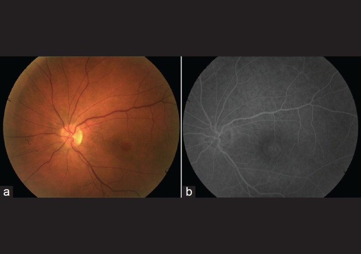
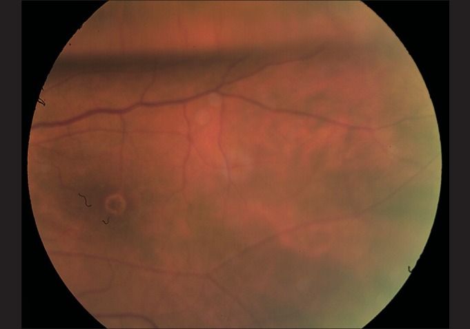
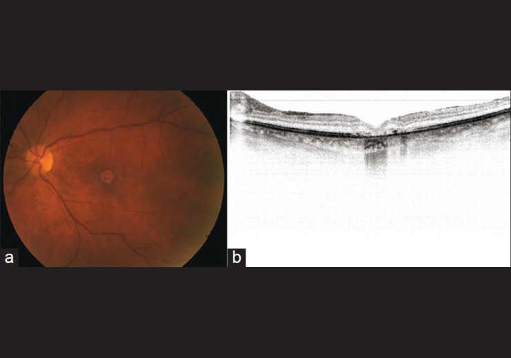
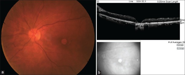
We report a case of progressive atrophy of the retinal pigment epithelium (RPE) after trypan-blue-assisted peeling of internal limiting membrane (ILM) for macular hole surgery. A 68-year-old Caucasian female underwent a 20-g pars plana vitrectomy for a chronic stage-3 macular hole. The ILM was stained with 0.06% trypan blue (VisionBlue™, DORC Netherlands) for 2 min after fluid air exchange. Dye was reapplied for another 2 min due to poor staining. The ILM was completely removed around the macular hole with forceps. RPE atrophy was noticed at the edge of the hole 1 month after surgery. It progressively increased in intensity and enlarged over 2 years. Her final visual acuity was counting fingers, significantly worse compared to her presenting visual acuity of 20/200. Progressive atrophy of RPE in our patient was most likely due to the toxicity of trypan blue. Reapplication of the dye may increase the likelihood of toxicity.
Conflict of interest statement
Figures
Similar articles
-
Retinal pigment epithelium atrophy after epiretinal membrane and internal limiting membrane peeling: case reports.Rom J Ophthalmol. 2022 Jan-Mar;66(1):79-83. doi: 10.22336/rjo.2022.16. Rom J Ophthalmol. 2022. PMID: 35531456 Free PMC article.
-
Atrophy of the retinal pigment epithelium following vitrectomy with trypan blue.Int Ophthalmol. 2009 Aug;29(4):239-41. doi: 10.1007/s10792-008-9195-5. Epub 2008 Apr 9. Int Ophthalmol. 2009. PMID: 18398579
-
Value of internal limiting membrane peeling in surgery for idiopathic macular hole and the correlation between function and retinal morphology.Acta Ophthalmol. 2009 Dec;87 Thesis 2:1-23. doi: 10.1111/j.1755-3768.2009.01777.x. Acta Ophthalmol. 2009. PMID: 19912135 Clinical Trial.
-
Indocyanine green-assisted peeling of the retinal internal limiting membrane during vitrectomy surgery for macular hole repair.Ophthalmology. 2001 Jul;108(7):1187-92. doi: 10.1016/s0161-6420(01)00593-0. Ophthalmology. 2001. PMID: 11425673
-
Retinal pigment epithelial changes after macular hole surgery with indocyanine green-assisted internal limiting membrane peeling.Am J Ophthalmol. 2002 Jan;133(1):89-94. doi: 10.1016/s0002-9394(01)01293-4. Am J Ophthalmol. 2002. PMID: 11755843
Cited by
-
A Review of Surgical Outcomes and Advances for Macular Holes.J Ophthalmol. 2018 Apr 18;2018:7389412. doi: 10.1155/2018/7389412. eCollection 2018. J Ophthalmol. 2018. PMID: 29850211 Free PMC article. Review.
-
Shortfalls of free autologous internal limiting membrane transplantation for highly myopic refractory macular holes in a long term follow-up.Graefes Arch Clin Exp Ophthalmol. 2024 Nov;262(11):3531-3541. doi: 10.1007/s00417-024-06533-7. Epub 2024 Jun 4. Graefes Arch Clin Exp Ophthalmol. 2024. PMID: 38832954 Free PMC article.
-
Macular Hole Surgery with Internal Limiting Membrane Peeling Facilitated by Membrane-Blue® versus Membrane-Blue-Dual®: A Retrospective Comparative Study.J Ophthalmol. 2016;2016:1292735. doi: 10.1155/2016/1292735. Epub 2016 Dec 5. J Ophthalmol. 2016. PMID: 28050275 Free PMC article.
-
Inner Retinal Layer and Outer Retinal Layer Findings after Macular Hole Surgery Assessed by means of Optical Coherence Tomography.J Ophthalmol. 2019 Apr 1;2019:3821479. doi: 10.1155/2019/3821479. eCollection 2019. J Ophthalmol. 2019. PMID: 31061725 Free PMC article. Review.
-
Retinal pigment epithelium atrophy after epiretinal membrane and internal limiting membrane peeling: case reports.Rom J Ophthalmol. 2022 Jan-Mar;66(1):79-83. doi: 10.22336/rjo.2022.16. Rom J Ophthalmol. 2022. PMID: 35531456 Free PMC article.
References
-
- Teba FA, Mohr A, Eckardt C, Wong D, Kusaka S, Joondeph BC, et al. Trypan blue staining in vitreoretinal surgery. Ophthalmology. 2003;110:2409–12. - PubMed
-
- Kodjikian L, Richter T, Halberstadt M, Beby F, Flueckiger F, Boehnke M, et al. Toxic effects of indocyanine green, infracyanine green, and trypan blue on the human retinal pigmented epithelium. Graefes Arch Clin Exp Ophthalmol. 2005;243:917–25. - PubMed
-
- Penha FM, Maia M, Eid Farah M, Príncipe AH, Freymüller EH, Maia A, et al. Effects of subretinal injections of indocyanine green, trypan blue, and glucose in rabbit eyes. Ophthalmology. 2007;114:899–908. - PubMed
-
- Schumann RG, Gandorfer A, Priglinger SG, Kampik A, Haritoglou C. Vital dyes for macular surgery: A comparative electron microscopy study of the internal limiting membrane. Retina. 2009;29:669–76. - PubMed
Publication types
MeSH terms
Substances
LinkOut - more resources
Full Text Sources
Other Literature Sources
Miscellaneous
