. 2013 Apr 25;2(3):e000080.
doi: 10.1161/JAHA.113.000080.

Arachidonic acid-induced dilation in human coronary arterioles: convergence of signaling mechanisms on endothelial TRPV4-mediated Ca2+ entry

Affiliations

Arachidonic acid-induced dilation in human coronary arterioles: convergence of signaling mechanisms on endothelial TRPV4-mediated Ca2+ entry

Xiaodong Zheng et al. J Am Heart Assoc. .

Abstract

Background: Arachidonic acid (AA) and/or its enzymatic metabolites are important lipid mediators contributing to endothelium-derived hyperpolarizing factor (EDHF)-mediated dilation in multiple vascular beds, including human coronary arterioles (HCAs). However, the mechanisms of action of these lipid mediators in endothelial cells (ECs) remain incompletely defined. In this study, we investigated the role of the transient receptor potential vanilloid 4 (TRPV4) channel in AA-induced endothelial Ca(2+) response and dilation of HCAs.

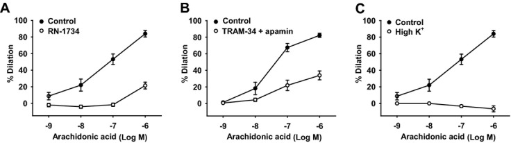
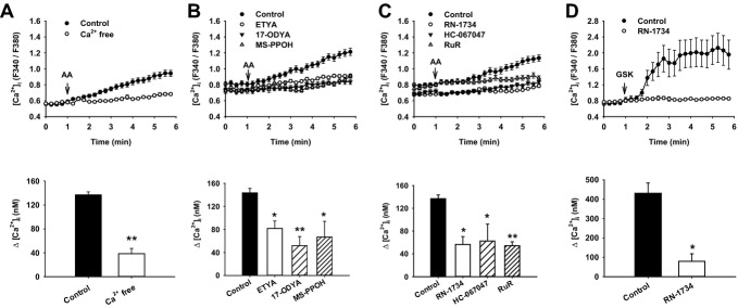
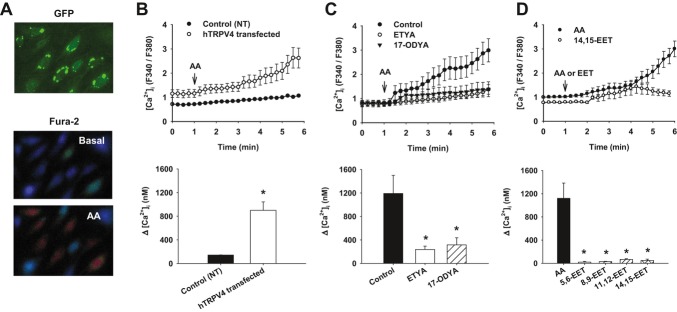
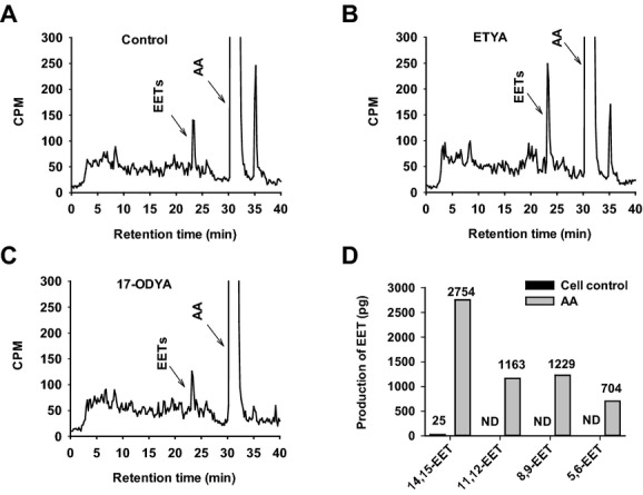
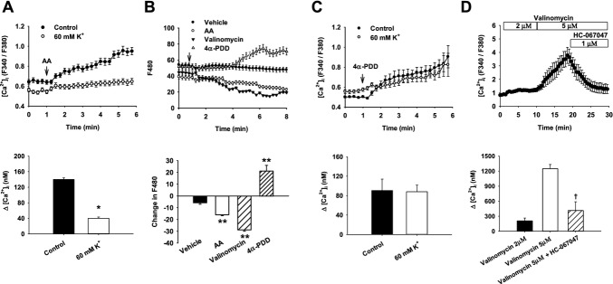
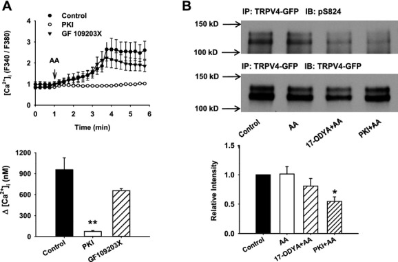
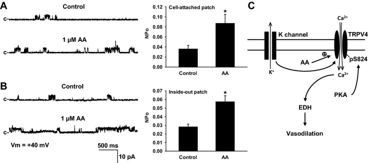
Methods and results: AA induced concentration-dependent dilation in isolated HCAs. The dilation was largely abolished by the TRPV4 antagonist RN-1734 and by inhibition of endothelial Ca(2+)-activated K(+) channels. In native and TRPV4-overexpressing human coronary artery ECs (HCAECs), AA increased intracellular Ca(2+) concentration ([Ca(2+)]i), which was mediated by TRPV4-dependent Ca(2+) entry. The AA-induced [Ca(2+)]i increase was inhibited by cytochrome P450 (CYP) inhibitors. Surprisingly, the CYP metabolites of AA, epoxyeicosatrienoic acids (EETs), were much less potent activators of TRPV4, and CYP inhibitors did not affect EET production in HCAECs. Apart from its effect on [Ca(2+)]i, AA induced endothelial hyperpolarization, and this effect was required for Ca(2+) entry through TRPV4. AA-induced and TRPV4-mediated Ca(2+) entry was also inhibited by the protein kinase A inhibitor PKI. TRPV4 exhibited a basal level of phosphorylation, which was inhibited by PKI. Patch-clamp studies indicated that AA activated TRPV4 single-channel currents in cell-attached and inside-out patches of HCAECs.

Conclusions: AA dilates HCAs through a novel mechanism involving endothelial TRPV4 channel-dependent Ca(2+) entry that requires endothelial hyperpolarization, PKA-mediated basal phosphorylation of TRPV4, and direct activation of TRPV4 channels by AA.


Figures

Figure 1.
Figure 1.
Role of TRPV4 and K+ channels in AA‐induced dilation of HCAs. A, AA dilated HCAs in a concentration‐dependent manner. The dilation was markedly inhibited by the TRPV4 antagonist RN‐1734 (20 μmol/L). B, AA‐induced dilation was inhibited by the combination of TRAM‐34 (1 μmol/L) and apamin (1 μmol/L), which selectively inhibit intermediate‐conductance KCa (IKCa) and small‐conductance KCa (SKCa) channels, respectively. C, AA‐induced dilation was eliminated by high extracellular K+ (80 mmol/L); n=5 to 7 patients/each group. *P<0.05 vs control. TRPV4 indicates transient receptor potential vanilloid 4; AA, arachidonic acid; HCA, human coronary artery.
Figure 2.
Figure 2.
AA‐induced increase of [Ca2+]i in native HCAECs. A, AA (3 μmol/L) increased [Ca2+]i, which was nearly eliminated in the absence of extracellular Ca2+ (Ca2+ free). B, AA‐elicited [Ca2+]i increase was inhibited by 3 CYP pathway inhibitors: ETYA (30 μmol/L), 17‐ODYA (10 μmol/L), and MS‐PPOH (30 μmol/L). C, Treatment of cells with TRPV4 antagonists RN‐1734 (20 μmol/L), HC‐067047 (1 μmol/L), and ruthenium red (RuR; 1 μmol/L) also markedly inhibited AA‐induced [Ca2+]i elevation. D, The TRPV4 agonist GSK1016790A (GSK; 10 nmol/L) increased [Ca2+]i, which was inhibited by RN‐1734. All data represent mean±SEM of ≥60 cells analyzed in 3 to 5 independent experiments. *P<0.05, **P<0.01 versus control. AA indicates arachidonic acid; ETYA, eicosatetraynoic acid; 17‐ODYA, 17‐octadecynoic acid; MS‐PPOH, N‐(methylsulfonyl)‐2‐(2‐propynyloxy)‐benzenehexanamide; HCAEC, human coronary artery endothelial cell; CYP, cytochrome P450; TRPV4, transient receptor potential vanilloid 4.
Figure 3.
Figure 3.
Ca2+ responses in hTRPV4‐overexpressing HCAECs. A, Representative images of endothelial cells expressing hTRPV4‐GFP (top) and corresponding cells loaded with fura‐2 before (middle) and after (bottom) AA treatment. B, Compared with nontransduced (NT) cells, AA (3 μmol/L) elicited augmented [Ca2+]i elevation in hTRPV4‐expressing cells. C, The AA‐induced Ca2+ response was inhibited by CYP pathway inhibitors ETYA (30 μmol/L) and 17‐ODYA (10 μmol/L). D, None of 4 isomeric EETs (3 or 10 μmol/L in some experiments) showed substantial activation of TRPV4 channels compared with AA. All data represent mean±SEM of ≥60 cells analyzed in 3 to 5 independent experiments. *P<0.05 versus control. hTRPV4 indicates human transient receptor potential vanilloid 4; HCAEC, human coronary artery endothelial cell; GFP, green fluorescent protein; AA, arachidonic acid; CYP, cytochrome P450; EET, epoxyeicosatrienoic acid.
Figure 4.
Figure 4.
Cytochrome P450 metabolism of AA in HCAECs. A, HCAECs were incubated with [14C]AA, metabolites of [14C]AA were extracted and resolved by reverse‐phase HPLC, and radioactivity of each fraction was determined by liquid scintillation spectrometry. Migration times of known standards are indicated. A major peak eluting at 22 to 24 minutes comigrated with the EET standard. This EET peak was not inhibited by CYP inhibitors ETYA (B; 30 μmol/L) or 17‐ODYA (C; 10 μmol/L). D, LC‐mass spectrometric analysis confirmed the production of 4 isomers of EETs— 14,15‐, 8,9‐, 11,12‐, and 5,6‐EET—in cells treated with AA (10 μmol/L). Endogenous EETs in nontreated cells were at low levels or not detectable (ND). CPM indicates counts per minute; AA, arachidonic acid; HCAEC, human coronary artery endothelial cell; HPLC, high‐performance liquid chromatography; EET, epoxyeicosatrienoic acid; CYP, cytochrome P450.
Figure 5.
Figure 5.
Modulation of TRPV4‐mediated Ca2+ influx by membrane potential. A, AA (3 μmol/L)‐elicited [Ca2+]i increase was blunted in the presence of 60 mmol/L extracellular K+. B, AA induced membrane hyperpolarization in HCAECs, as indicated by a decrease in PMPI fluorescence intensity (F480). In contrast, the TRPV4 agonist 4α‐PDD (3 μmol/L) depolarized the membrane potential. Valinomycin (K+‐selective ionophore, 2 μmol/L) was used as a positive control to indicate membrane hyperpolarization. C, The TRPV4 specific agonist 4α‐PDD (5 μmol/L) increased [Ca2+]i in HCAECs, and the increase was not affected by 60 mmol/L K+. D, Membrane hyperpolarization by valinomycin resulted in Ca2+ influx in hTRPV4‐expressing HCAECs, which was inhibited by HC‐067047. The data represent mean±SEM of ≥60 cells analyzed in 3 to 5 independent experiments. *P<0.05 vs control, **P<0.01 vs vehicle, P<0.05 vs valinomycin (5 μmol/L). TRPV4 indicates transient receptor potential vanilloid 4; AA, arachidonic acid; 4α‐PDD, 4α‐phorbol‐12,13‐didecanoate; HCAEC, human coronary artery endothelial cell; PMPI, plasma membrane potential indicator.
Figure 6.
Figure 6.
Regulation of TRPV4 activation by protein phosphorylation. A, In hTRPV4‐expressing HCAECs, the AA‐induced [Ca2+]i increase was almost abolished by the PKA inhibitor PKI (1 μmol/L), whereas this AA response was only partially but not significantly attenuated by the PKC inhibitor GF 109203X (1 μmol/L). B, TRPV4 is phosphorylated at serine‐824 in unstimulated HCAECs expressing the hTRPV4‐GFP fusion protein. The level of serine‐824 phosphorylation was not further increased by AA (3 μmol/L). PKI (1 μmol/L) significantly inhibited serine‐824 phosphorylation. Endothelial cells were treated with the indicated reagents, and TRPV4‐GFP was immunoprecipitated with GFP antibodies and detected with pS824 antibodies (upper panel). The same membrane was reprobed with GFP antibodies to detect total TRPV4 protein (middle panel). All data represent mean±SEM from 3 to 5 independent experiments. At least 60 cells analyzed in (A). *P<0.05, **P<0.01 vs control. TRPV4 indicates transient receptor potential vanilloid 4; HCAEC, human coronary artery endothelial cell; AA, arachidonic acid; PKA, protein kinase A; PKI, protein kinase A inhibitor; IP, immunoprecipitation; IB, immunoblotting; GFP, green fluorescent protein.
Figure 7.
Figure 7.
Single‐channel properties of hTRPV4 overexpressed in HCAECs. A, TRPV4 single‐channel currents (left) were recorded from a cell‐attached patch at the indicated membrane potentials (Vm). Right panel depicts average current–voltage relationship for single‐channel currents, with a calculated slope conductance determined over voltage ranges of 0 to 60 and −40 to 0 mV, respectively. Data are mean±SE from 5 membrane patches. B, Single‐channel activity of TRPV4 (left) was low under basal conditions but markedly increased following bath perfusion of the TRPV4 agonist 4α‐PDD (1 μmol/L). Shown on the right are the corresponding amplitude histograms in relation to open‐state probability (NPo). Data are representative of >5 membrane patches. Dashed lines indicate current level when the channel is closed. Cell‐attached recordings were obtained with a normal extracellular solution (140 Na+, 5 Cs+) in the pipette and a high‐K+ (140 K+) bath solution to zero the cell membrane potential. TRPV4 indicates transient receptor potential vanilloid 4; HCAEC, human coronary artery endothelial cell.
Figure 8.
Figure 8.
Effect of AA on hTRPV4 single‐channel currents in HCAECs. AA (1 μmol/L) increased TRPV4 single‐channel currents in both cell‐attached patches (A) and inside‐out patches, which lacked intracellular constituents (B). Left, representative recordings at the indicated membrane potential (Vm). Note that c is the current level when the channel is closed. Right, summary of channel open‐state probability (NPo). Cell‐attached and inside‐out patch recordings before and after treatment with AA were performed with a normal extracellular solution (140 Na+,5 Cs+) in the pipette and a high‐K+ (140 K+) solution in the bath. n=6 patches/each group. *P<0.05 vs control. C, Possible mechanism for AA‐induced Ca2+ entry through TRPV4 channels in human coronary endothelial cells and subsequent dilation of coronary arterioles. AA indicates arachidonic acid; TRPV4, transient receptor potential vanilloid 4; HCAEC, human coronary artery endothelial cell; PKA, protein kinase A; EDH, endothelium‐dependent hyperpolarization.

Similar articles

Cited by

References

    1. Edwards G, Feletou M, Weston AH. Endothelium‐derived hyperpolarising factors and associated pathways: a synopsis. Pflugers Arch. 2010; 459:863-879 - PubMed
    1. Campbell WB, Fleming I. Epoxyeicosatrienoic acids and endothelium‐dependent responses. Pflugers Arch. 2010; 459:881-895 - PMC - PubMed
    1. Shimokawa H. Hydrogen peroxide as an endothelium‐derived hyperpolarizing factor. Pflugers Arch. 2010; 459:915-922 - PubMed
    1. Miura H, Bosnjak JJ, Ning G, Saito T, Miura M, Gutterman DD. Role for hydrogen peroxide in flow‐induced dilation of human coronary arterioles. Circ Res. 2003; 92:e31-e40 - PubMed
    1. Liu Y, Bubolz AH, Mendoza S, Zhang DX, Gutterman DD. H2O2 is the transferrable factor mediating flow‐induced dilation in human coronary arterioles. Circ Res. 2011; 108:566-573 - PMC - PubMed

Publication types

MeSH terms

LinkOut - more resources