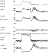
Physiology-based MR imaging assessment of CSF flow at the foramen magnum with a valsalva maneuver
- PMID: 23620074
- PMCID: PMC7965609
- DOI: 10.3174/ajnr.A3509
Physiology-based MR imaging assessment of CSF flow at the foramen magnum with a valsalva maneuver
Abstract
Background and purpose: MR imaging is currently not used to evaluate CSF flow changes due to short-lasting physiological maneuvers. The purpose of this study was to evaluate the ability of MR imaging to assess the CSF flow response to a Valsalva maneuver in healthy participants.
Materials and methods: A cardiac-gated fast cine-PC sequence with ≤15-second acquisition time was used to assess CSF flow in 8 healthy participants at the foramen magnum at rest, during, and immediately after a controlled Valsalva maneuver. CSF mean displacement volume VCSF during the cardiac cycle and CSF flow waveform App were determined. A work-in-progress real-time pencil-beam imaging method with temporal resolution ≤56 ms was used to scan 2 participants for 90 seconds during which resting, Valsalva, and post-Valsalva CSF flow, respiration, and HR were continuously recorded. Results were qualitatively compared with invasive craniospinal differential pressure measurements from the literature.
Results: Both methods showed 1) a decrease from baseline in VCSF and App during Valsalva and 2) an increase in VCSF and App immediately after Valsalva compared with values measured both at rest and during Valsalva. Whereas fast cine-PC produced a single CSF flow waveform that is an average over many cardiac cycles, pencil-beam imaging depicted waveforms for each heartbeat and was able to capture many dynamic features of CSF flow, including transients synchronized with the Valsalva maneuver.
Conclusions: Both fast cine-PC and pencil-beam imaging demonstrated expected changes in CSF flow with Valsalva maneuver in healthy participants. The real-time capability of pencil-beam imaging may be necessary to detect Valsalva-related transient CSF flow obstruction in patients with pathologic conditions such as Chiari I malformation.
Figures


References
-
- Williams B. Simultaneous cerebral and spinal fluid pressure recordings. I. Technique, physiology, and normal results. Acta Neurochir (Wien) 1981;58:167–85 - PubMed
-
- Heiss JD, Patronas N, DeVroom HL, et al. Elucidating the pathophysiology of syringomyelia. J Neurosurg 1999;91:553–62 - PubMed
-
- Sansur CA, Heiss JD, DeVroom HL, et al. Pathophysiology of headache associated with cough in patients with Chiari I malformation. J Neurosurg 2003;98:453–58 - PubMed
-
- Tachibana S, Iida H, Yada K. Significance of positive Queckenstedt test in patients with syringomyelia associated with Arnold-Chiari malformations. J Neurosurg 1992;76:67–71 - PubMed
Publication types
MeSH terms
LinkOut - more resources
Full Text Sources
Other Literature Sources
Medical
