. 2013 Sep 1;22(17):3397-414.
doi: 10.1093/hmg/ddt190. Epub 2013 Apr 24.

Missense mutation in mouse GALC mimics human gene defect and offers new insights into Krabbe disease

Affiliations

Missense mutation in mouse GALC mimics human gene defect and offers new insights into Krabbe disease

Gregory B Potter et al. Hum Mol Genet. .

Abstract

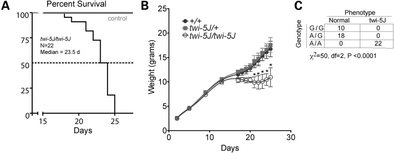
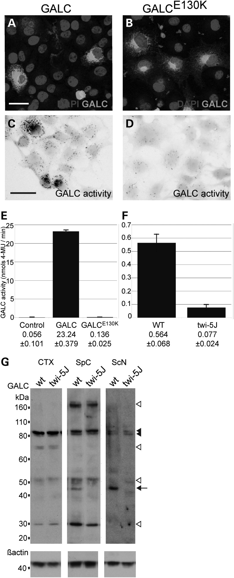
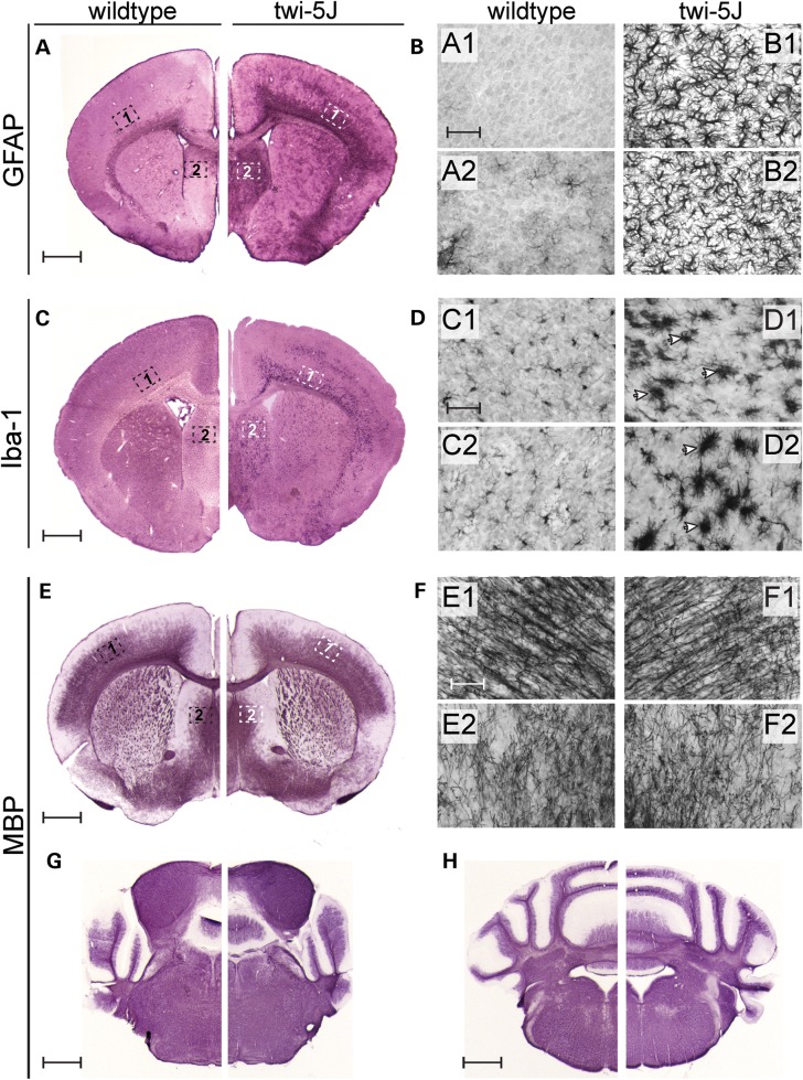
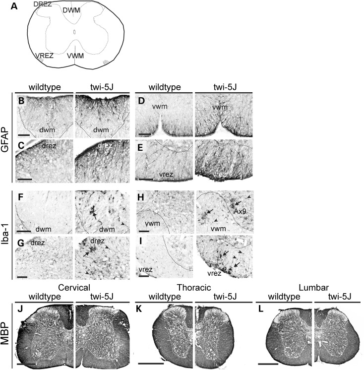
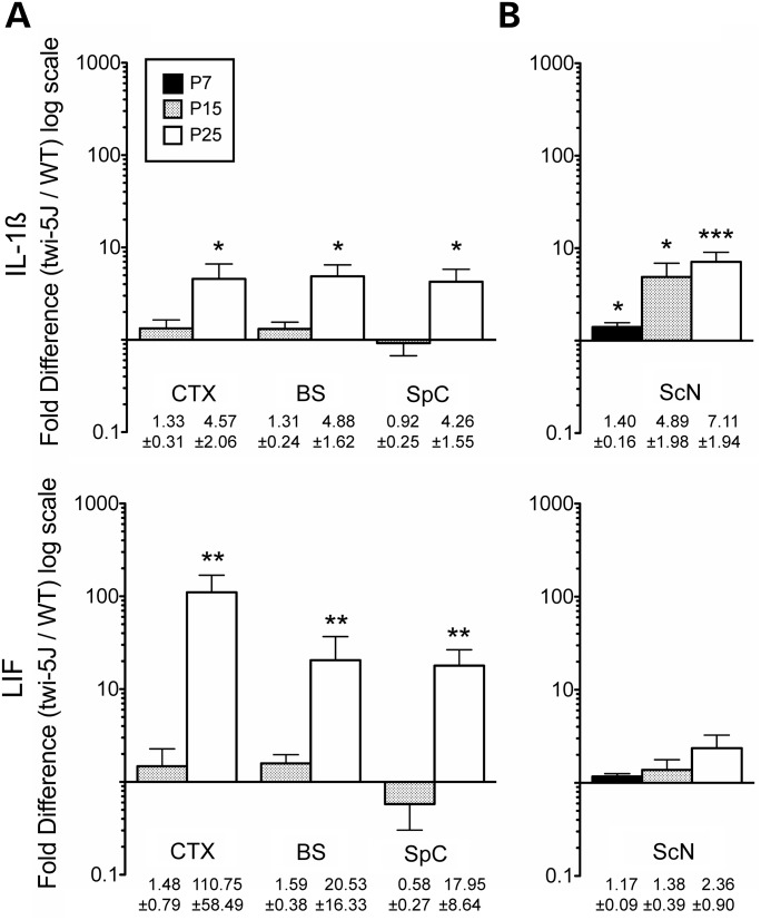
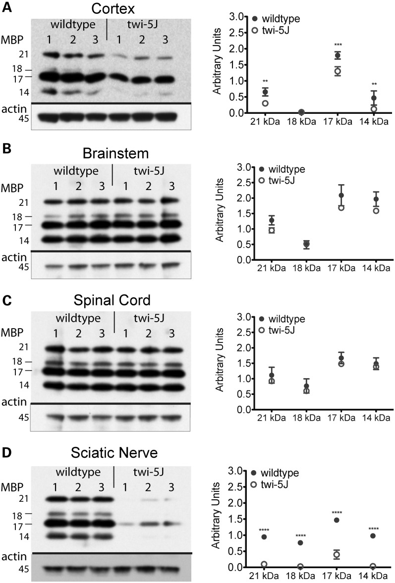
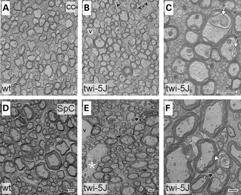
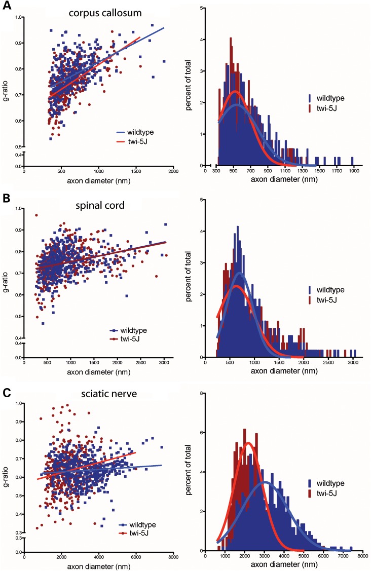
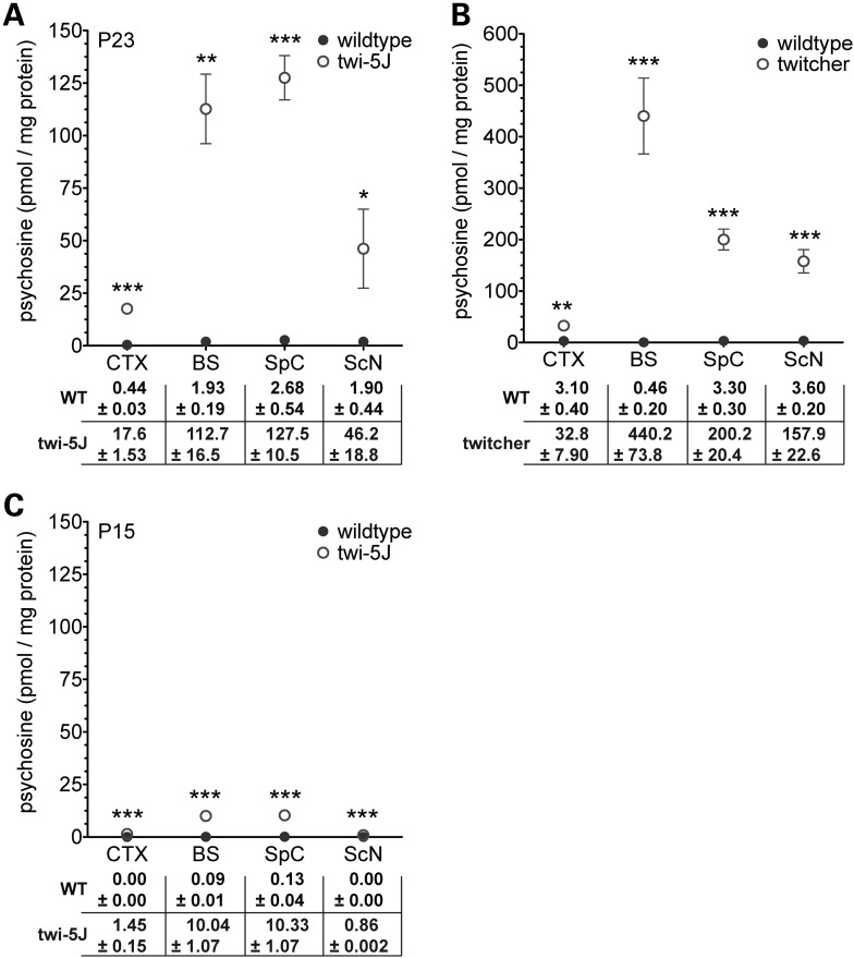
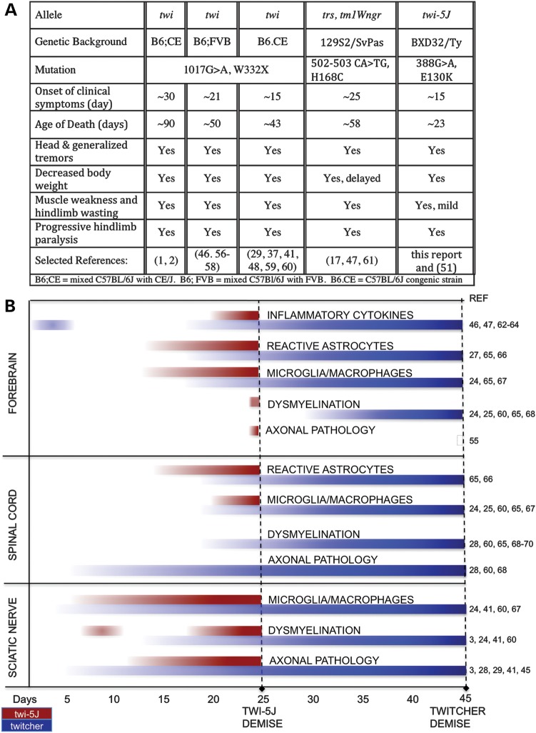
Krabbe disease is a devastating pediatric leukodystrophy caused by mutations in the galactocerebrosidase (GALC) gene. A significant subset of the infantile form of the disease is due to missense mutations that result in aberrant protein production. The currently used mouse model, twitcher, has a nonsense mutation not found in Krabbe patients, although it is similar to the human 30 kb deletion in abrogating GALC expression. Here, we identify a spontaneous mutation in GALC, GALCtwi-5J, that precisely matches the E130K missense mutation in patients with infantile Krabbe disease. GALCtwi-5J homozygotes show loss of enzymatic activity despite normal levels of precursor protein, and manifest a more severe phenotype than twitcher, with half the life span. Although neuropathological hallmarks such as gliosis, globoid cells and psychosine accumulation are present throughout the nervous system, the CNS does not manifest significant demyelination. In contrast, the PNS is severely hypomyelinated and lacks large diameter axons, suggesting primary dysmyelination, rather than a demyelinating process. Our data indicate that early demise is due to mechanisms other than myelin loss and support an important role for neuroinflammation in Krabbe disease progression. Furthermore, our results argue against a causative relationship between psychosine accumulation, white matter loss and gliosis.


Figures

Figure 1.
Figure 1.
A missense mutation in Galc associates with the twi-5J phenotype. (A) Kaplan–Meier survival analysis of twi-5J. The median survival was 23.5 days (n = 22). (B) Weight curve of twi-5J. Homozygous GALCtwi-5J mice weighed significantly less than heterozygotes for GALCtwi-5J and wild-type mice after P20. *P < 0.01. (C) Mice homozygous for 388A are twi-5J. Genotype analysis using allele-specific PCR to GALC nucleotide 388 compared with phenotype. Wild-type (G/G) and heterozygous (G/A) mice are unaffected, while all homozygous (A/A) mice show neurological symptoms and are phenotypic twi-5J.
Figure 2.
Figure 2.
The E130K substitution in GALC abolishes GALC enzymatic activity without affecting precursor protein levels in vivo. (AD) COS-7 cells were transfected with an expression construct for GALC (A and C) or GALCE130K (B and D). (A and B) GALC (white) was detected by immunofluorescence using a GALC-specific antibody. (C and D) GALC activity (black) was visualized using a GALC histochemical assay. Cell bodies are false-colored grey. Scale bar in A, C = 50 µm. (E) GALC activity (nmols/min) was measured from 15 µg of COS-7 cells extracts transfected with expression constructs for wild-type GALC or GALCE130K. Controls are mock-transfected cells. (E) GALC activity (nmols/min) measured from 15 µg of whole brain extracts from WT and twi-5J. Activity is presented as mean ± SD. (G) Western blot analysis using an affinity purified GALC antibody on extracts from wild-type (wt) and twi-5J collected from cortex (CTX), spinal cord (SpC) and sciatic nerve (ScN). Expression of ∼80, ∼50 and ∼30 kDa GALC bands are equivalent in cortical extracts between wild-type and twi-5J (P > 0.05). In contrast, a ∼45 kDa band is absent in extracts collected from mutant spinal cord and sciatic nerve compared with wild-type controls (P < 0.001) (arrow). The ∼80 kDa GALC precursor (black arrowheads) is present in all tissues. White arrowheads point to GALC species differentially regulated between tissues. Blots were re-probed using a beta-actin antibody to control for protein loading. Apparent molecular mass is indicated on the left. Extracts from three independent wild-type and twi-5J animals were analyzed with equivalent results; a representative immunoblot is shown.
Figure 3.
Figure 3.
Twi-5J display gliosis and globoid cell accumulation in the forebrain. (A and B) GFAP immunohistochemistry on coronal sections from P25 wild-type (A) and twi-5J (B) indicate an increase in reactive astrocytes within the CNS. Higher magnification views of the cortex and septum in wild-type (A1–A2) and twi-5J (B1–B2) show the presence of large, multi-branched and densely stained cells. (C and D) Iba-1 immunohistochemistry on coronal sections from P25 wild-type (C) and twi-5J (D) reveals the presence of large macrophages (arrowheads) in the mutant cortex and striatum. (EH) MBP immunohistochemistry on coronal sections from P25 wild-type and twi-5J reveal comparable myelination in the CNS. Higher magnification views of the cortex and striatum in wild-type (E1–E2) and twi-5J (F1–F2). Scale bars: A–H = 1 mm; A1–F1, A2–F2 = 50 µm.
Figure 4.
Figure 4.
Spinal cords of twi-5J exhibit gliosis and macrophage accumulation. Cross-sections of spinal cords from wild-type and twi-5J were immunolabeled for GFAP (BE), Iba-1 (FI) and MBP (J–L). (A) Schematic of cross-section through spinal cord with regions of interest annotated. (B–E) Reactive astrocytes, marked by changes in astrocyte morphology and increased GFAP expression, are present in twi-5J (right panels, white arrowheads) in the dorsal (DWM) and ventral white matter (VWM) and near the dorsal root entry (drez) and ventral root exit zones (vrez). (F–I) Iba-1 immunolabeling reveals large Iba-1+ cells consistent with macrophages (arrows) and an increase in small Iba-1+ microglia (arrowhead) in ventral and dorsal white matter of twi-5J spinal cord. Ax9, lamina 9 (axial muscle innervation). (J–L) MBP expression is similar between wild-type (left) and twi-5J (right) at all levels of the spinal cord. Scale bars: B–I = 100 µm; J–L = 500 µm.
Figure 5.
Figure 5.
Inflammatory cytokine expression is increased in twi-5J nervous system. Quantitative RT–PCR for IL1-β and LIF expression was performed on RNA samples processed from the cortex (CTX), brainstem (BS), spinal cord (SpC) and sciatic nerve (ScN) of wild-type and twi-5J at P7, P15 or P25 (mean ± SEM, n = 3). (A) IL1-β and LIF expression is significantly increased in the CNS at P25, but not at P15. (B) In the sciatic nerve, IL1-β expression is significantly elevated and progressively increases starting at P7, while LIF expression is not increased in twi-5J compared with wild-type. *P < 0.05; **P < 0.01; ***P < 0.005.
Figure 6.
Figure 6.
Expression of MBP isoforms is significantly decreased in twi-5J. Protein samples from three post-natal day 25 wild-type and twi-5J animals were separated by SDS–PAGE and examined for MBP expression by immunoblot analysis. MBP isoform expression was significantly decreased in the cortex (A) and sciatic nerve (D) of twi-5J. MBP expression within the brainstem and spinal cord were not significantly different. Each band is quantified relative to beta-actin expression and presented in arbitrary units. Data is plotted on the right as mean ± SD. ****P < 0.0001; ***P < 0.001; **P < 0.01.
Figure 7.
Figure 7.
Electron microscopy of corpus callosum and spinal cord reveals ultrastructural abnormalities in twi-5J. Electron micrographs of the corpus callosum (cc) (AC) and spinal cord (SpC) (DF) post-natal day 25 wild-type (A and D) and twi-5J (B and C, E and F). Large diameter axons within the corpus callosum of twi-5J are normally myelinated. There is a small increase in unmyelinated medium-caliber axons (arrowheads) and lipid inclusions (white arrow, C). Spinal cord of twi-5J manifests occasional electron-light regions representing early signs of axonal loss (white asterisk), degenerating axons surrounded by compact myelin (white arrowhead) and lipid inclusions within axons (black arrow). v, blood vessel. Scale bar = 1 µm.
Figure 8.
Figure 8.
Analysis of myelinated axons reveals perturbations in axonal populations in twi-5J. G-ratio plot (left panels) and myelinated axon diameter distribution (right panels) for control and twi-5J in the corpus callosum (A), spinal cord (B) and sciatic nerve (C). (A) In the corpus callosum, a subset of small caliber axons (330–400 nm) are hypermyelinated and the number of large diameter axons (>650 nm) are reduced by ∼20% in twi-5J. (B) No significant changes in g-ratio or axon diameter are detected in the spinal cord. (C) Twi-5J exhibit loss of large axons (>4000 nm) and hypomyelination of small (900–2000 nm) and medium axons (2000–4000 nm). Significantly greater hyper-myelination of axons < 3000 nm in twi-5J offsets hypomyelination to generate averaged g-ratios comparable to wild-type at these axon diameters.
Figure 9.
Figure 9.
The sciatic nerves of twi-5J are severely hypomyelinated with macrophage accumulation. (AD) MBP immunohistochemistry on cross-sections (A and B) and transverse-sections (C and D) through the sciatic nerve of P23 wild-type (A and C) and twi-5J (B and D) indicate significant myelin-deficiency in the PNS. (E and F) Toluidine blue staining on thin sections through the sciatic nerve of P25 wild-type control (E) and twi-5J (F). Compared with wild-type, the twi-5J sciatic nerve is less compact, exhibits ∼22% loss of axons and contain numerous hypomyelinated axons (arrowheads), macrophages (M) and large lipid-filled vesicles (arrows). (G and H) Iba-1 immunohistochemistry on cross-sections reveal accumulation of globoid cells (arrows) in twi-5J. Iba-1 also detects non-reactive macrophages in control and twi-5J sciatic nerve (arrowhead). (IK) Electron microscopy of sciatic nerve of twi-5J reveals numerous macrophages, hypomyelinated axons and lipid inclusions compared with wild-type. Inset in J shows globoid cell. Black Arrows point to lipid inclusions. Arrowheads indicate hypomyelinated axons. M, macrophage; RB, Remak bundle. Scale bars: A–D = 250 µm; E–H = 25 µm; I–K = 1 µm.
Figure 10.
Figure 10.
Psychosine levels are increased throughout the nervous system of twi-5J. (A) Psychosine concentrations (pmol/mg protein) were measured in tissue samples collected from cortex (CTX), brainstem (BS), spinal cord (SpC) and sciatic nerve (ScN) of post-natal day 23 twi-5J (n = 3) and wild-type littermates (n = 3). (B) Psychosine concentrations measured from P23–P25 twitcher and wild-type littermate tissue samples (n = 3–5). (C) Psychosine concentrations measured in tissue samples collected from cortex, brainstem, spinal cord and sciatic nerve of post-natal day 15 twi-5J (n = 3) and wild-type littermates (n = 3). Data plotted as mean ± SEM. ***P < 0.001; **P < 0.01; *P < 0.05.
Figure 11.
Figure 11.
Comparison of twi-5J and twitcher clinical symptoms and disease progression. (A) Clinical symptoms of Krabbe disease mouse models. (B) Schematic comparing characteristics of disease progression between twi-5J and twitcher in the CNS and PNS. In the forebrain, axonal pathology is observed in twi-5J but not in twitcher. In contrast, significant axonal pathology is not detected in the spinal cord of twi-5J. The twi-5J sciatic nerve exhibits early perturbation in myelination, normalization of g-ratios and MBP expression by P15, and severe dysmyelination by P25. Inflammatory cytokine progression in the spinal cord and sciatic nerve has not been examined in twitcher (56,70). A comparison of psychosine accumulation is presented in Figure 10.

References

    1. Igisu H., Suzuki K. Progressive accumulation of toxic metabolite in a genetic leukodystrophy. Science. 1984;224:753–755. doi:10.1126/science.6719111. - DOI - PubMed
    1. Xu Y.H., Barnes S., Sun Y., Grabowski G.A. Multi-system disorders of glycosphingolipid and ganglioside metabolism. J. Lipid Res. 2010;51:1643–1675. doi:10.1194/jlr.R003996. - DOI - PMC - PubMed
    1. Jacobs J.M., Scaravilli F., De Aranda F.T. The pathogenesis of globoid cell leucodystrophy in peripheral nerve of the mouse mutant twitcher. J. Neurol. Sci. 1982;55:285–304. doi:10.1016/0022-510X(82)90127-7. - DOI - PubMed
    1. Suzuki K., Taniike M. Murine model of genetic demyelinating disease: the twitcher mouse. Microsc. Res. Tech. 1995;32:204–214. doi:10.1002/jemt.1070320304. - DOI - PubMed
    1. Wenger D.A. 1997. Krabbe disease. GeneReviews at GeneTests: Medical Genetics Information Resource (database online). Copyright, University of Washington, Seattle, 2010.

Publication types

MeSH terms