. 2013 Dec;7(4):320-6.
doi: 10.1007/s12105-013-0444-1. Epub 2013 Apr 26.

Papillary variant of squamous cell carcinoma arising on the gingiva: 61 cases reported from within a larger series of gingival squamous cell carcinoma

Affiliations

Papillary variant of squamous cell carcinoma arising on the gingiva: 61 cases reported from within a larger series of gingival squamous cell carcinoma

Sarah G Fitzpatrick et al. Head Neck Pathol. 2013 Dec.

Abstract

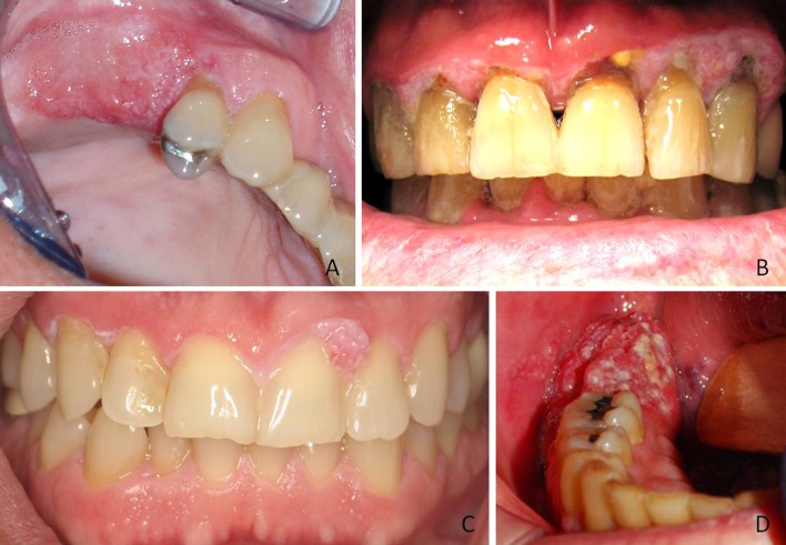
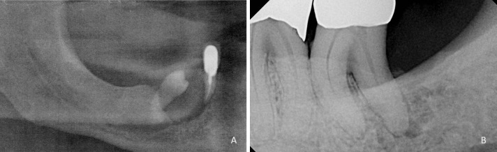
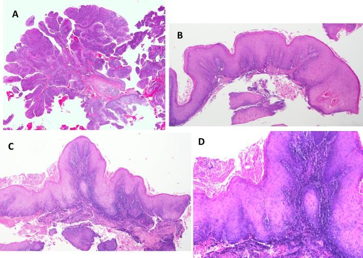
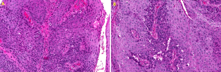
Papillary squamous cell carcinoma (PSCCA) is a rarely occurring variant of squamous cell carcinoma (SCCA) with distinctive exophytic and papillary features and a more favorable prognosis than conventional SCCA. The larynx is the most commonly affected site in the head and neck. The oral cavity, oropharynx, sinonasal tract, and nasopharynx are also affected. Within the oral cavity cases have been reported on the alveolar ridge, oral mucosa, floor of the mouth, ventral tongue, and rarely other areas. We identified 61 cases of gingival PSCCA within the parameters of a larger study of 519 cases of gingival SCCA. We evaluated the clinical and histologic features of these lesions. The average age of the PSCCA patient was 74 years, with a very slight male predominance of 1.2:1. The mandible was affected nearly twice as often (64 %, n = 39) as the maxilla (35 %, n = 21, and 1 % of cases unspecified), and the most common location by far was the mandibular posterior region (52 %, n = 32). Most lesions were reportedly present over 2 months in duration (48 %, n = 29) prior to biopsy but a significant amount of clinicians were unsure of the duration of the lesion as well (36 %, n = 22). Only 10 cases (16 %) were reportedly present less than 2 months. The most common clinical presentation was that of an erythematous or mixed white and red exophytic mass (74 %, n = 45). 62 % (n = 38) of submitting clinicians considered a malignant or premalignant lesion in their differential diagnosis, but other clinical impressions included papillomas, reactive gingival lesions, and fungal infections. Histologically, 88 % (n = 52 of 59 cases graded) of the lesions were either well or moderately-well differentiated. PSCCA is a rare subtype of SCCA which has been reported infrequently involving the gingiva or alveolar ridges but should be considered by clinicians for the differential diagnosis of papillary gingival masses.


Figures

Fig. 1
Fig. 1
Clinical presentation of PSCCA. a Pebbly raised red and white lesion involving the alveolar ridge distal to tooth #5 (Photo courtesy Dr. Tom Kassube). b Diffuse mixed lesion with papillary areas “cuffing” the facial gingival margins of the maxillary anterior teeth (Photo courtesy Dr. Douglas Johnson). c Thin white leukoplakia of marginal gingiva from teeth #6–11 with area of PSCCA interproximally between teeth #9–10 (Photo courtesy Dr. Brian Van Aelst). d Exophytic red and white mass on the alveolar ridge distal to tooth #30 (Photo courtesy Dr. James Heit)
Fig. 2
Fig. 2
Radiographic presentation of PSCCA. a Bone loss and displacement of the mandibular right premolar in a patient with PSCCA (Radiograph courtesy Dr. Eric Fox). b Irregular radiolucency surrounding the distal root of tooth #18 mimicking a lesion of endodontic-periodontic origin (Radiograph courtesy Dr. Luis Rosario)
Fig. 3
Fig. 3
Histologic features of PSCCA. a Low power histomicrograph demonstrating the prominent papillary nature of neoplastic proliferation surrounding fibrovascular cores and minimal surface keratinization (H&E, ×2 magnification). b Low power features demonstrating the verrucoid papillary nature with bulbous rete ridges with keratin pearl formation from a different case. (H&E, ×4 magnification). c Papillary proliferation with elongated and bulbous rete ridge formation. (H&E, ×4 magnification). d Higher magnification of case in image c demonstrating individual cell keratinization and keratin pearl formation. (H&E, ×10 magnification)
Fig. 4
Fig. 4
a Higher magnification demonstrating abundant atypical features in the proliferating epithelium including increased abnormal mitoses with increased nuclear/cytoplasmic ratios, crowding of cells and pleomorphism. (H&E, ×20 magnification). b Higher magnification demonstrating increased mitoses, enlarged and abnormal nuclei and numerous dyskeratotic cells (H&E, ×20 magnification)

References

    1. Crissman JD, Kessis T, Shah KV, Fu YS, Stoler MH, Zarbo RJ, Weiss MA. Squamous papillary neoplasia of the adult upper aero digestive tract. Hum Pathol. 1988;19:1387–1396. doi: 10.1016/S0046-8177(88)80231-4. - DOI - PubMed
    1. Thompson LDR, Wenig BM, Heffner DK, Gnepp DR. Exophytic and papillary squamous cell carcinomas of the larynx: a clinicopathologic series of 104 cases. Otolaryngol Head Neck Surg. 1999;120:718–724. doi: 10.1053/hn.1999.v120.a92773. - DOI - PubMed
    1. Russell JO, Hoschar AP, Scharpf J. Papillary squamous cell carcinoma of the head and neck: a clinicopathologic series. Am J Otolaryngol. 2011;32:557–563. doi: 10.1016/j.amjoto.2010.09.010. - DOI - PubMed
    1. Cobo F, Talavera P, Concha A. Relationship of human papillomavirus with papillary squamous cell carcinoma of the upper aero digestive tract: a review. Int J Surg Pathol. 2008;16(2):127–136. doi: 10.1177/1066896908314700. - DOI - PubMed
    1. Ishiyama A, Eversole LR, Ross DA, Raz Y, Kerner MM, Yao S, Blackwell K, Feneberg R, Bell TS, Calcaterra TC. Papillary squamous neoplasms of the head and neck. Laryngosope. 1994;104(12):1446–1452. - PubMed

LinkOut - more resources