. 2013 Aug;31(8):1283-92.
doi: 10.1002/jor.22348. Epub 2013 Apr 25.

Effects of cartilage impact with and without fracture on chondrocyte viability and the release of inflammatory markers

Affiliations

Effects of cartilage impact with and without fracture on chondrocyte viability and the release of inflammatory markers

Josef A Stolberg-Stolberg et al. J Orthop Res. 2013 Aug.

Abstract

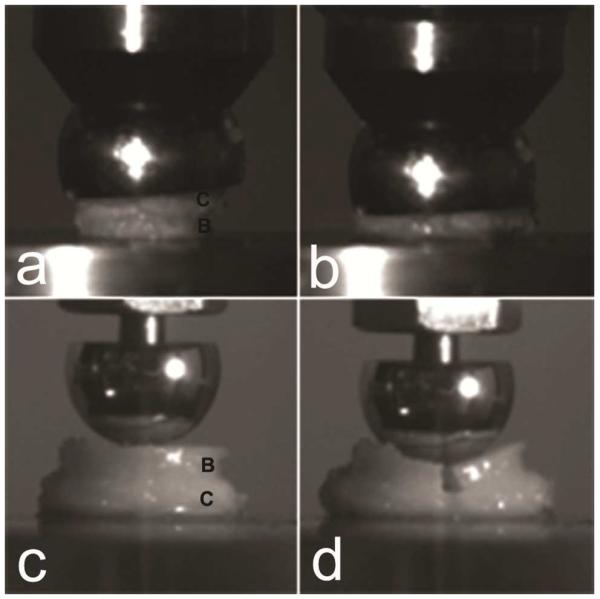
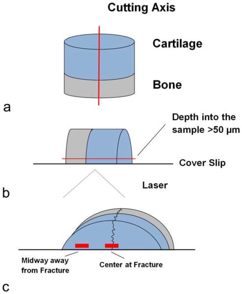
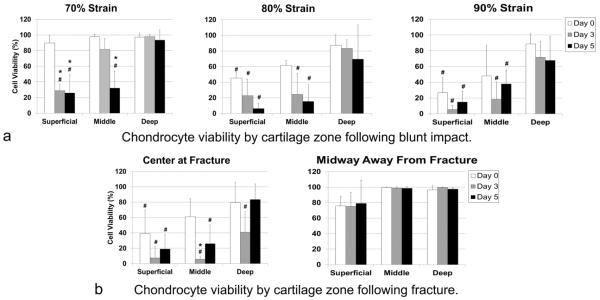
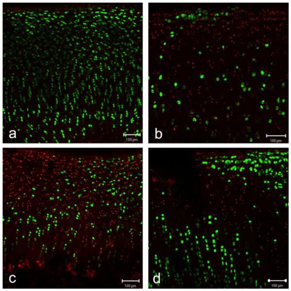
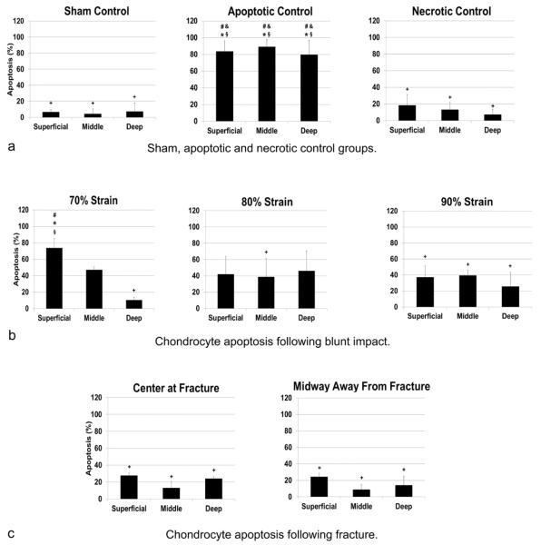
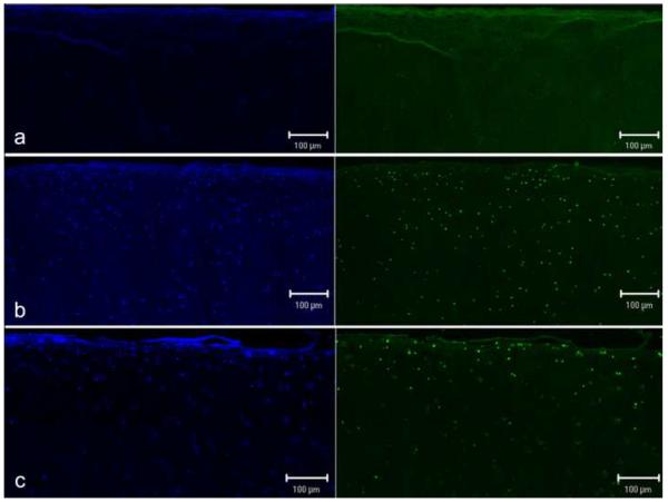
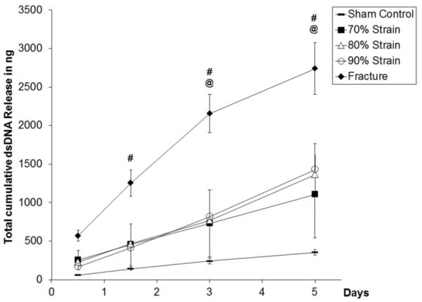
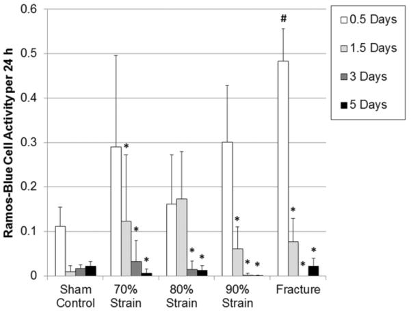
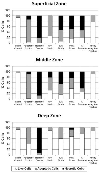
Post-traumatic arthritis (PTA) frequently develops after intra-articular fracture of weight bearing joints. Loss of cartilage viability and post-injury inflammation have both been implicated as possible contributing factors to PTA progression. To further investigate chondrocyte response to impact and fracture, we developed a blunt impact model applying 70%, 80%, or 90% surface-to-surface compressive strain with or without induction of an articular fracture in a cartilage explant model. Following mechanical loading, chondrocyte viability, and apoptosis were assessed. Culture media were evaluated for the release of double-stranded DNA (dsDNA) and immunostimulatory activity via nuclear factor kappa B (NF-κB) activity in Toll-like receptor (TLR) -expressing Ramos-Blue reporter cells. High compressive strains, with or without articular fracture, resulted in significantly reduced chondrocyte viability. Blunt impact at 70% strain induced a loss in viability over time through a combination of apoptosis and necrosis, whereas blunt impact above 80% strain caused predominantly necrosis. In the fracture model, a high level of primarily necrotic chondrocyte death occurred along the fracture edges. At sites away from the fracture, viability was not significantly different than controls. Interestingly, both dsDNA release and NF-κB activity in Ramos-Blue cells increased with blunt impact, but was only significantly increased in the media from fractured cores. This study indicates that the mechanism of trauma determines the type of chondrocyte death and the potential for post-injury inflammation.


Figures

Figure 1
Figure 1
Loading of osteochondral cores (C = cartilage, B = bone). For the blunt impact model (a) a hemisphere was placed with the flat face on the cartilage surface. The concave adapter attached to the loadframe allowed self-alignement and completely blunt loading (b). For the fracture model (c) a truncated ball in contact with bone caused sample frature (d).
Figure 2
Figure 2
Osteochondral cores were dissected perpendicular to the surface (a). Confocal images were taken > 50 μm into the tissue (b). Viability of blunt impacted explants was assessed at the center of the core. Cell death of fractured cores was measured at the fracture edge and at the midway between fracture and outer edge (c).
Figure 3
Figure 3
Chondrocyte viability by cartilage zone following (a) blunt impact and (b) fracture. A significant decrease of viability over time could only be detected after 70% strain in the superficial and middle zone. (#indicates statistically significant differences from sham controls; *indicates statistically significant differences from day 0).
Figure 4
Figure 4
Live/ Dead assay confocal microscope images. 70% strain blunt impact caused a significant loss of viability over time from (a) day 0 to (b) day 5. No time dependent cell death could be detected after (c) blunt impact ≥ 80% strain or (d) at fracture edges.
Figure 5
Figure 5
Apoptosis as detected by ISOL at Day 5. Only the superficial layer of 70% strain samples showed a significant difference to sham, necrotic controls and midway of fractured samples (p≤0.027) (#indicates statistically significant differences from sham controls; *indicates statistically significant differences from necrosis; + indicates statistically significant differences from apoptosis, & indicates statistically significant difference from fractured edge, § indicates statistically significant difference from midway between fracture and outer edge).
Figure 6
Figure 6
Apoptotic Cell death was confirmed by ISOL. The blue channel on the left shows all nuclei within the section. The green channel on the right shows apoptotic nuclei. Only very few positively stained chondrocytes could be found in the necrotic control samples (a). The apoptotic control samples treated with staurosporine (b) showed similar fluorescence as the superficial layer of samples loaded with 70% strain blunt impact (c).
Figure 7
Figure 7
Cumulative dsDNA release (ng) in media as normalized to cartilage height. Fractured samples showed significantly higher dsDNA release than sham controls at days 1.5, 3 and 5 and bluntly loaded samples at days 3 and 5. (#indicates statistically significant differences from sham controls, @ indicates statistically significant differences from all bluntly loaded samples).
Figure 8
Figure 8
Ramos Blue Cell Activity per 24 h of culture time and normalized to cartilage height. There was a peak of immunostimulatory activity in all treated groups at day 0.5. Only the fractured group showed a significant difference to sham controls. (#indicates statistically significant differences from sham controls; *indicates statistically significant differences from day 0).
Figure 9
Figure 9
Relative percentage of live, apoptotic, and necrotic cells at day 5. 70% low-strain blunt impact lead to predominantly apoptosis whereas 80% and greater high-strain blunt impact and fracture caused predominantly necrosis in addition to apoptosis.

Similar articles

Cited by

References

    1. Brown TD, Johnston RC, Saltzman CL, Marsh JL, Buckwalter JA. Posttraumatic osteoarthritis: a first estimate of incidence, prevalence, and burden of disease. J Orthop Trauma. 2006 Nov-Dec;20(10):739–744. - PubMed
    1. Matta JM. Fractures of the acetabulum: accuracy of reduction and clinical results in patients managed operatively within three weeks after the injury. J Bone Joint Surg Am. 1996 Nov;78(11):1632–1645. - PubMed
    1. Stufkens SA, Knupp M, Horisberger M, Lampert C, Hintermann B. Cartilage lesions and the development of osteoarthritis after internal fixation of ankle fractures: a prospective study. J Bone Joint Surg Am. 2010 Feb;92(2):279–286. - PubMed
    1. Anderson DD, Chubinskaya S, Guilak F, et al. Post-traumatic osteoarthritis: improved understanding and opportunities for early intervention. J Orthop Res. 2011 Jun;29(6):802–809. - PMC - PubMed
    1. Olson SA, Guilak F. From articular fracture to posttraumatic arthritis: A black box that needs to be opened. Journal of Orthopaedic Trauma. 2006 Nov-Dec;20(10):661–662. - PubMed

Publication types

MeSH terms