. 2013 Aug;94(Pt 8):1749-1760.
doi: 10.1099/vir.0.052910-0. Epub 2013 Apr 25.

MERS-coronavirus replication induces severe in vitro cytopathology and is strongly inhibited by cyclosporin A or interferon-α treatment

Affiliations

MERS-coronavirus replication induces severe in vitro cytopathology and is strongly inhibited by cyclosporin A or interferon-α treatment

Adriaan H de Wilde et al. J Gen Virol. 2013 Aug.

Abstract

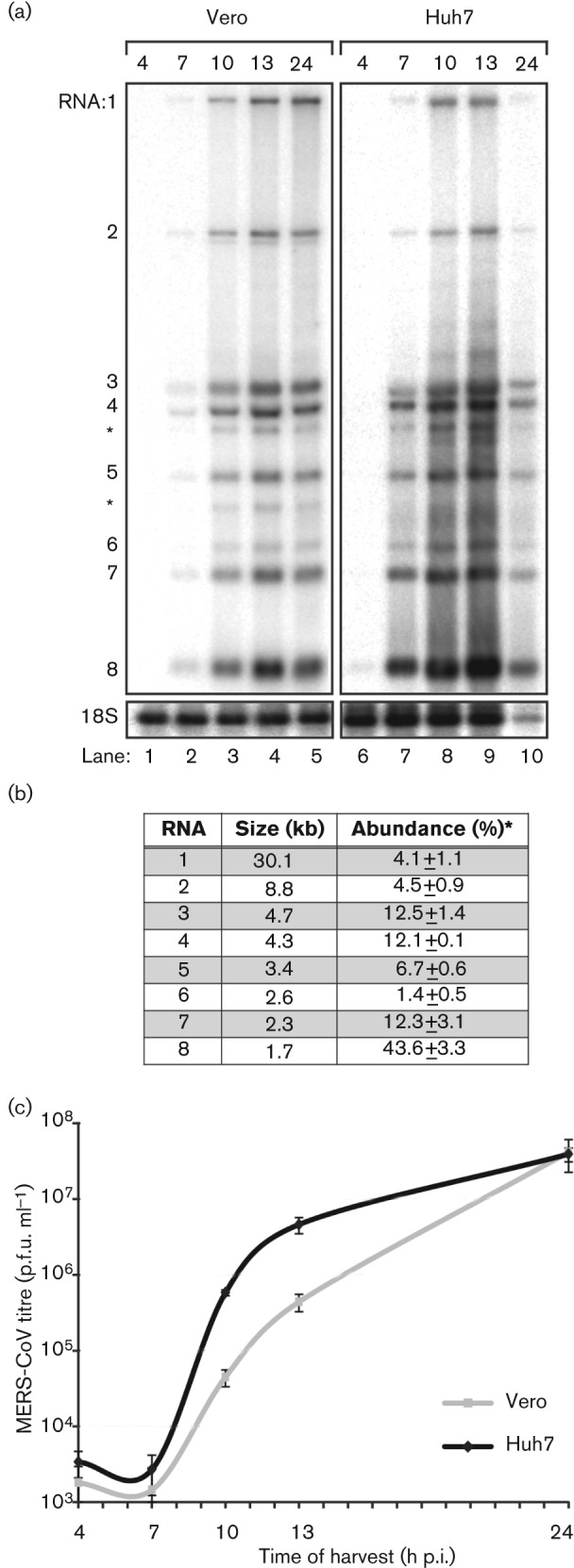
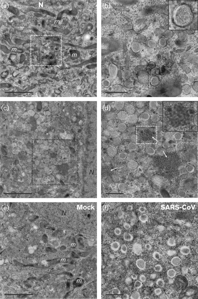
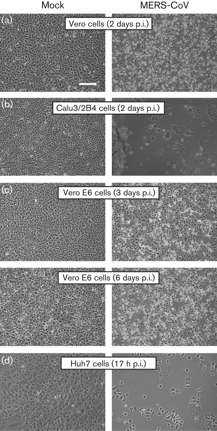
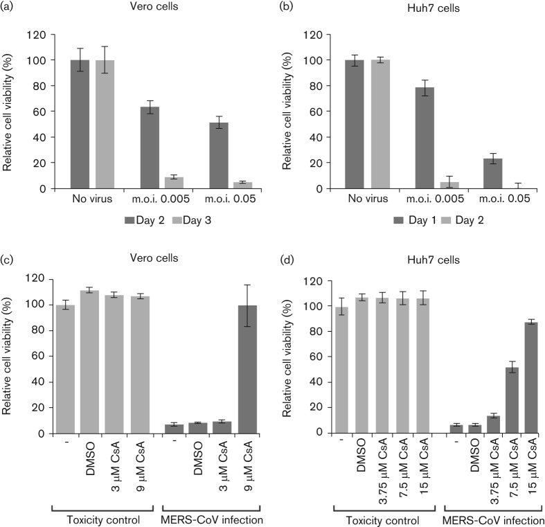
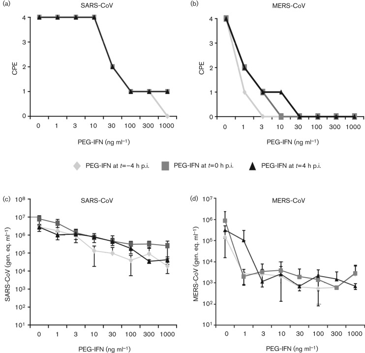
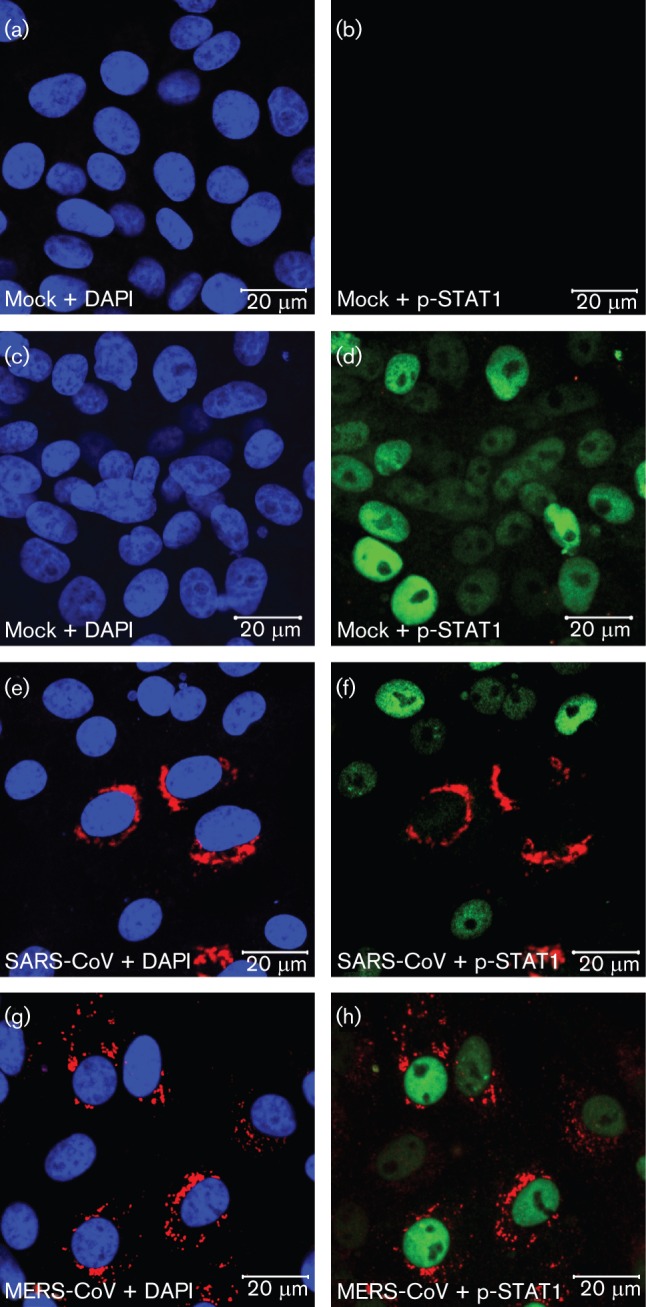
Coronavirus (CoV) infections are commonly associated with respiratory and enteric disease in humans and animals. The 2003 outbreak of severe acute respiratory syndrome (SARS) highlighted the potentially lethal consequences of CoV-induced disease in humans. In 2012, a novel CoV (Middle East Respiratory Syndrome coronavirus; MERS-CoV) emerged, causing 49 human cases thus far, of which 23 had a fatal outcome. In this study, we characterized MERS-CoV replication and cytotoxicity in human and monkey cell lines. Electron microscopy of infected Vero cells revealed extensive membrane rearrangements, including the formation of double-membrane vesicles and convoluted membranes, which have been implicated previously in the RNA synthesis of SARS-CoV and other CoVs. Following infection, we observed rapidly increasing viral RNA synthesis and release of high titres of infectious progeny, followed by a pronounced cytopathology. These characteristics were used to develop an assay for antiviral compound screening in 96-well format, which was used to identify cyclosporin A as an inhibitor of MERS-CoV replication in cell culture. Furthermore, MERS-CoV was found to be 50-100 times more sensitive to alpha interferon (IFN-α) treatment than SARS-CoV, an observation that may have important implications for the treatment of MERS-CoV-infected patients. MERS-CoV infection did not prevent the IFN-induced nuclear translocation of phosphorylated STAT1, in contrast to infection with SARS-CoV where this block inhibits the expression of antiviral genes. These findings highlight relevant differences between these distantly related zoonotic CoVs in terms of their interaction with and evasion of the cellular innate immune response.


Figures

Fig. 1.
Fig. 1.
Kinetics of MERS-CoV replication in Vero and Huh7 cells. Vero and Huh7 cells were infected with MERS-CoV (m.o.i. of 5). (a) Hybridization analysis of viral mRNAs isolated from MERS-CoV-infected cells using an oligonucleotide recognizing the viral genome and all sg mRNAs. Additional minor bands of ~3 and ~4 kb were observed (*) and may represent additional viral mRNA species that remain to be studied in more detail. However, the corresponding positions in the ORF4a/b and ORF5 coding regions do not contain a canonical core TRS sequence (AACGAA; van Boheemen et al., 2012) that might provide a direct explanation for their synthesis. (b) Analysis of the relative molarities of viral genome and each of the sg mRNAs (% of total viral mRNA). mRNA sizes were calculated on the basis of the TRS positions in the viral genome sequence (van Boheemen et al., 2012). Phosphorimager quantification was performed on the gel lanes with the RNA samples isolated from Vero cells at 10, 13 and 24 h p.i. (Fig. 1a; lanes 3–5, respectively; mean±sd). (c) Release of infectious MERS-CoV progeny into the medium of infected Vero or Huh7 cells at the indicated time points, as determined by plaque assay (mean±sd; n = 4).
Fig. 2.
Fig. 2.
Selected rabbit antisera raised against SARS-CoV and mouse hepatitis virus (MHV) nsps cross-react with MERS-CoV proteins. (a) MERS-CoV-infected Vero cells (m.o.i. of 5) were fixed at 8 h p.i. For immunofluorescence microscopy, cells were double-labelled with a mouse mAb recognizing dsRNA (bottom row) and rabbit antisera raised against SARS-CoV nsp3, nsp4, nsp5 or nsp8, or MHV nsp4 (top row). Bar, 20 µm. (b) Sequence comparison of the C-terminal domain of nsp4 of SARS-CoV (isolate Frankfurt 1), MERS-CoV (strain EMC/2012) and MHV (strain A59). The SARS-CoV and MHV sequences correspond to the synthetic peptides used to raise rabbit anti-nsp4 sera. Residues conserved in all three viruses are highlighted in yellow, whereas residues conserved in two out of three are highlighted in grey. Amino acid numbers refer to the full-length pp1a sequence. (c) Monolayers of Vero, Vero E6, Huh7 and Calu3/2B4 cells were infected with MERS-CoV (m.o.i. of 5) and double-labelled for dsRNA (green) and nsp3 (red). Bar, 40 µm.
Fig. 3.
Fig. 3.
Membrane structures induced by MERS-CoV infection. (a–d) Electron micrographs of thin sections (100 nm) of MERS-CoV-infected Vero cells at 8 h p.i. Low magnification images of a cell containing a small cluster of DMVs (a), enlarged in (b). Some DMVs are indicated by black arrowheads and the inset displays a higher magnification of the boxed DMV in (b). Extensive membrane alterations in the perinuclear region are shown in (c), with the boxed area displayed at higher magnification in (d), where CMs (white arrows, inset) embedded in clusters of DMVs (black arrowheads) can be observed. (e, f) For comparison, (e) shows the unaltered cytoplasm of a mock-infected cell and (f) contains SARS-CoV-induced DMV (black arrowheads) as observed after HPF and freeze substitution. N, nucleus; m, mitochondria. Bars, 2 µm (a, c, e); 500 nm (b, d, f).
Fig. 4.
Fig. 4.
MERS-CoV infection induces severe cytopathology in monkey and human cell lines. Monolayers of Vero (a), Calu3/2B4 (b), Vero E6 (c) and Huh7 (d) cells were infected with MERS-CoV (m.o.i. of 0.05) and analysed by light microscopy at the indicated time points. Bar, 100 µm.
Fig. 5.
Fig. 5.
Development of an assay to screen for compounds inhibiting MERS-CoV replication. Vero (a, c) and Huh7 (b, d) cells in a 96-well plate format were infected at an m.o.i. of 0.005 or 0.05. Mock-infected cells (no virus) were used as a reference for unchanged cell viability (their relative viability was set at 100 %). Infected Vero cells were incubated for 2 (dark shading) or 3 (light shading) days (a) and Huh7 cells were incubated for 1 (dark shading) or 2 (light shading) days (b). (c) Vero cells were infected (dark shading) or not (light shading) with MERS-CoV (m.o.i. of 0.005) in the presence of 3 or 9 µM CsA, or 0.09 % DMSO as a solvent control. (d) Huh7 cells were infected (dark shading) or not (light shading) with MERS-CoV (m.o.i. of 0.005) in the presence of 3.75, 7.5 or 15 µM CsA, or 0.15 % DMSO. The graphs in (c) and (d) show the results of a representative experiment (mean±sd; n = 4). All experiments were repeated at least twice.
Fig. 6.
Fig. 6.
Sensitivity of MERS-CoV and SARS-CoV to PEG-IFN. Vero cells were incubated with 0–1000 ng PEG-IFN ml−1 at t = −4, t = 0 and t = 4 h p.i. Cells were infected with 100 TCID50 virus per well (a, b). At 2 days p.i., cells were examined for CPE. The effect of PEG-IFN treatment on CPE induced by SARS-CoV (a) or MERS-CoV (b) is shown. CPE was scored as none (0), mild (1), moderate (2), severe (3) or complete (4). (c, d) Viral genomes in the culture medium of virus-infected cells were determined by RT-PCR. The influence of PEG-IFN treatment on the viral RNA load [genome equivalents (gen. eq.) ml−1] in the supernatants of cells infected with SARS-CoV (c) or MERS-CoV (d) is shown.
Fig. 7.
Fig. 7.
IFN-α induced nuclear translocation of p-STAT1 in MERS-CoV-infected Vero cells. Confocal immunofluorescence microscopy of uninfected Vero cells (a–d) and Vero cells infected (m.o.i. of 1) with SARS-CoV (e, f) or MERS-CoV (g, h). At 8 h p.i. cells were left untreated (a, b) or treated (c–h) with 1000 ng PEG-IFN ml−1 for 30 min, fixed and double-labelled with antisera against SARS-CoV nsp3 (red; a–h), or p-STAT1 (green; b, d, f, h), and nuclear DNA was stained with DAPI (blue; a, c, e, g).

References

    1. Belouzard S., Millet J. K., Licitra B. N., Whittaker G. R. (2012). Mechanisms of coronavirus cell entry mediated by the viral spike protein. Viruses 4, 1011–1033 10.3390/v4061011 - DOI - PMC - PubMed
    1. Bergman S. J., Ferguson M. C., Santanello C. (2011). Interferons as therapeutic agents for infectious diseases. Infect Dis Clin North Am 25, 819–834 10.1016/j.idc.2011.07.008 - DOI - PMC - PubMed
    1. Brockway S. M., Clay C. T., Lu X. T., Denison M. R. (2003). Characterization of the expression, intracellular localization, and replication complex association of the putative mouse hepatitis virus RNA-dependent RNA polymerase. J Virol 77, 10515–10527 10.1128/JVI.77.19.10515-10527.2003 - DOI - PMC - PubMed
    1. Cameron M. J., Kelvin A. A., Leon A. J., Cameron C. M., Ran L., Xu L., Chu Y. K., Danesh A., Fang Y. & other authors (2012). Lack of innate interferon responses during SARS coronavirus infection in a vaccination and reinfection ferret model. PLoS ONE 7, e45842 10.1371/journal.pone.0045842 - DOI - PMC - PubMed
    1. Cervantes-Barragan L., Züst R., Weber F., Spiegel M., Lang K. S., Akira S., Thiel V., Ludewig B. (2007). Control of coronavirus infection through plasmacytoid dendritic-cell-derived type I interferon. Blood 109, 1131–1137 10.1182/blood-2006-05-023770 - DOI - PMC - PubMed

Publication types