Case Reports
. 2013 Jul;24(8):1305-12.
doi: 10.1681/ASN.2012090913. Epub 2013 Apr 25.

Nephrotic syndrome caused by immune-mediated acquired LCAT deficiency

Affiliations
Case Reports

Nephrotic syndrome caused by immune-mediated acquired LCAT deficiency

Satoshi Takahashi et al. J Am Soc Nephrol. 2013 Jul.

Abstract

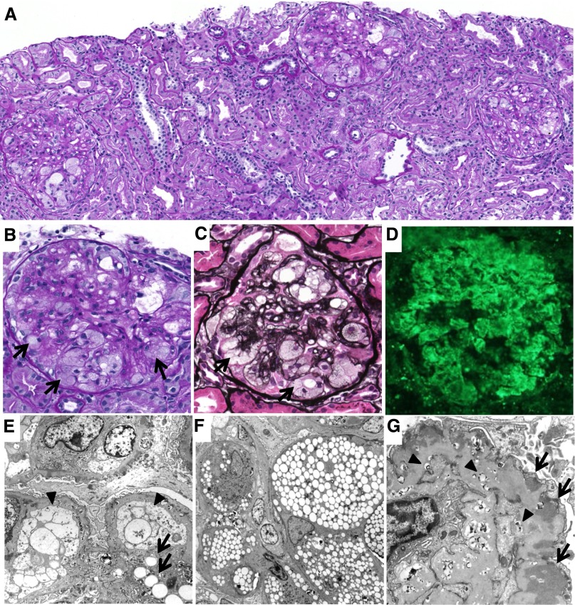
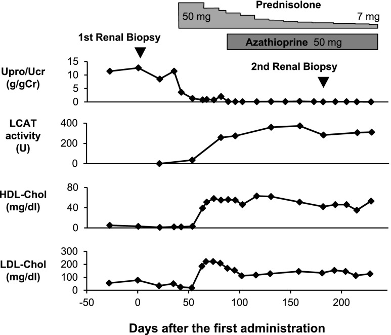
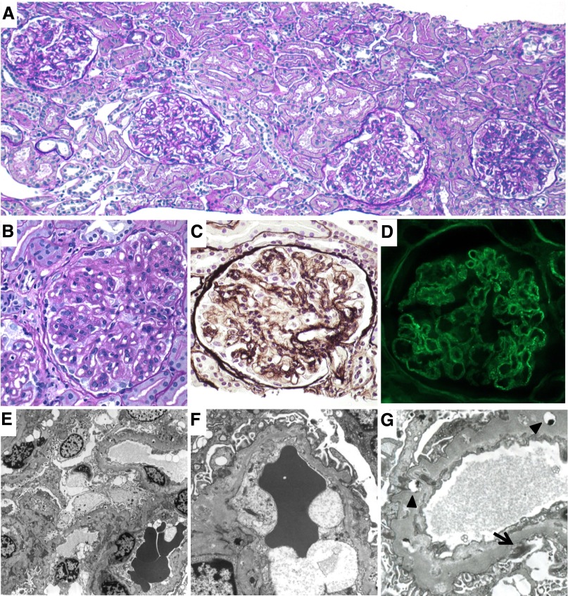
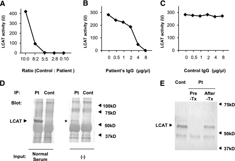
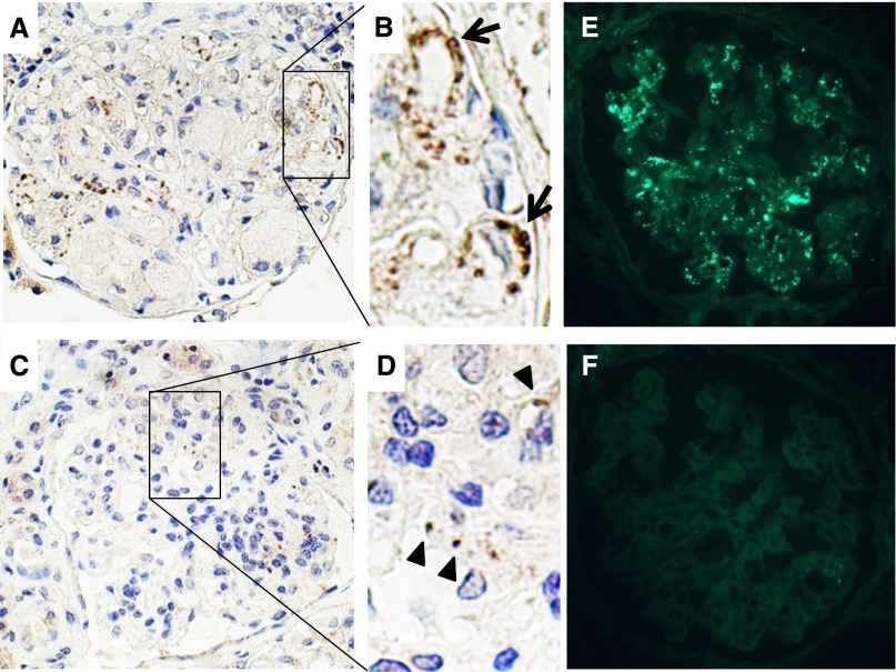
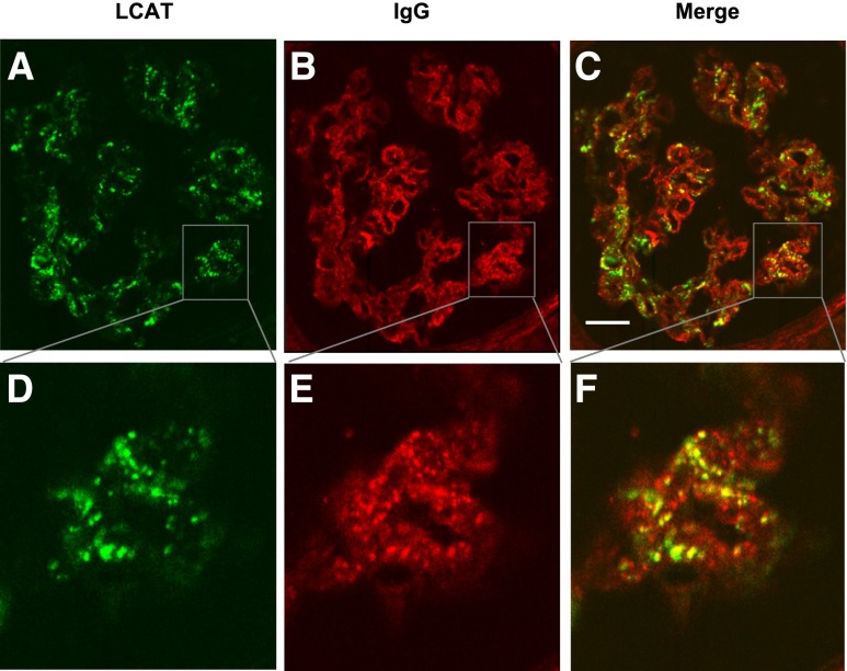
Lecithin-cholesterol acyltransferase (LCAT) is an enzyme involved in maintaining cholesterol homeostasis. In familial LCAT deficiency (FLD), abnormal lipid deposition causes renal injury and nephrotic syndrome, frequently progressing to ESRD. Here, we describe a 63-year-old Japanese woman with no family history of renal disease who presented with nephrotic syndrome. The laboratory data revealed an extremely low level of serum HDL and undetectable serum LCAT activity. Renal biopsy showed glomerular lipid deposition with prominent accumulation of foam cells, similar to the histologic findings of FLD. In addition, she had subepithelial electron-dense deposits compatible with membranous nephropathy, which are not typical of FLD. A mixing test and coimmunoprecipitation study demonstrated the presence of an inhibitory anti-LCAT antibody in the patient's serum. Immunohistochemistry and immunofluorescence detected LCAT along parts of the glomerular capillary walls, suggesting that LCAT was an antigen responsible for the membranous nephropathy. Treatment with steroids resulted in complete remission of the nephrotic syndrome, normalization of serum LCAT activity and HDL level, and disappearance of foam cell accumulation in renal tissue. In summary, inhibitory anti-LCAT antibody can lead to glomerular lesions similar to those observed in FLD.


Figures

Figure 1.
Figure 1.
Histologic findings at the first renal biopsy. (A–C) Light microscopy findings. Glomeruli show marked foam cell accumulation (arrows), together with mild mesangial proliferation and expansion (periodic acid–Schiff staining in A and B, periodic acid silver-methenamine staining in C). (D) Immunofluorescence findings. Coarse granular depositions of IgG are observed mainly along the capillary walls. (E–G) Electron microscopy findings. (E) Lipid deposits are observed in the mesangial area (arrows). The endothelial cells become enlarged and occlude the capillary lumina (arrowheads). (F) The foam cells are accumulated in the capillary lumina. (G) Irregular thickening of the GBM with subepithelial electron-dense deposits (arrows) and osmiophilic lipid deposits (arrowheads) are also observed. Original magnification, ×100 in A and D; ×400 in B and C; ×1200 in E; ×1000 in F; ×2500 in G.
Figure 2.
Figure 2.
Clinical course of the patient. Soon after the initiation of prednisolone therapy, the urinary protein/urinary creatinine ratio decreases and complete remission of the nephrotic syndrome is achieved. The severely deficient LCAT activity and markedly decreased HDL cholesterol level increase to within normal range after the initiation of the prednisolone therapy. Upro/Ucr, urinary protein/urinary creatinine ratio; HDL-Chol, HDL cholesterol; LDL-Chol, LDL cholesterol.
Figure 3.
Figure 3.
Histologic findings at the second renal biopsy. (A–C) Light microscopy findings. Marked reduction of the foam cells and improvement of the mesangial lesions (periodic acid-Schiff staining in A and B; periodic acid silver-methenamine staining in C). (D) Immunofluorescence findings. Weak fine granular depositions of IgG are observed along the capillary walls. (E–G) Electron microscopy findings. (E and F) The foam cells disappear from the capillary lumina. (G) The apparent decrease in the amount of subepithelial electron-dense deposits (arrow), although the GBM show diffuse thickening with fine osmiophilic lipid depositions (arrowheads). Original magnification, ×100 in A; ×400 in B–D; ×1200 in E; ×4000 in F and G.
Figure 4.
Figure 4.
Inhibitory autoantibody against LCAT. (A) Mixing test conducted using the patient’s serum and healthy control serum shows the existence of a factor inhibitory for LCAT activity in the patient’s serum. (B) Addition of the purified patient’s IgG to healthy control serum decreases the LCAT activity in a dose-dependent manner. (C) Addition of purified IgG from healthy control serum to a different control serum has no effect on the LCAT activity. (D) Coimmunoprecipitation study. Patient’s serum (Pt) or healthy control serum (Cont) is preincubated with protein G beads, and then incubated with a different control serum (normal serum). LCAT of normal serum is immunoprecipitated with the patient’s serum (arrowhead), but not with control serum. In addition, protein G beads preincubated with the patient’s serum alone capture LCAT (asterisk), demonstrating that the patient’s serum contained immune complexes composed of IgG and LCAT. (E) Western blot analysis for LCAT. Healthy control serum and the patient’s serum before steroid therapy (Pre-Tx) and after steroid therapy (After-Tx) are pretreated with albumin and IgG removal kits. The treated sera are then subjected to Western blotting for LCAT using rabbit monoclonal anti-LCAT antibody. LCAT is detected in both healthy control serum and the patient’s serum after, but not before, the steroid treatment (arrowhead).
Figure 5.
Figure 5.
Immunostaining for LCAT. (A–D) Immunohistochemical staining on formalin-fixed, paraffin-embedded renal biopsy specimens with rabbit monoclonal anti-LCAT antibody. (A and B) Coarse granular staining is detected mainly along the glomerular capillary walls (arrows) in the first renal biopsy sample. (C and D) Faint granular staining (arrowheads) is observed in the second renal biopsy sample 5 months after the treatment. (E and F) Immunofluorescence on frozen sections of the second renal biopsy. (E) Coarse granular staining is mainly observed along the glomerular capillary walls by rabbit monoclonal anti-LCAT antibody. (F) Specific staining is not observed after elimination of the first antibody.
Figure 6.
Figure 6.
Double immunostaining for LCAT and IgG on frozen sections of the second renal biopsy by confocal microscopy. (A and D) LCAT is detected along the glomerular capillary walls by rabbit monoclonal anti-LCAT antibody (green). (B and E) IgG deposition is also detected along the capillary walls by anti-human IgG (red). (C and F) Partial colocalization of LCAT and IgG is observed along the capillary walls (yellow). Scale bar, 20 μm.

References

    1. Jonas A: Lecithin cholesterol acyltransferase. Biochim Biophys Acta 1529: 245–256, 2000 - PubMed
    1. Zannis VI, Chroni A, Krieger M: Role of apoA-I, ABCA1, LCAT, and SR-BI in the biogenesis of HDL. J Mol Med (Berl) 84: 276–294, 2006 - PubMed
    1. Calabresi L, Franceschini G: Lecithin:cholesterol acyltransferase, high-density lipoproteins, and atheroprotection in humans. Trends Cardiovasc Med 20: 50–53, 2010 - PubMed
    1. Gjone E, Norum KR: Familial serum cholesterol ester deficiency. Clinical study of a patient with a new syndrome. Acta Med Scand 183: 107–112, 1968 - PubMed
    1. Carlson LA, Philipson B: Fish-eye disease. A new familial condition with massive corneal opacities and dyslipoproteinaemia. Lancet 2: 922–924, 1979 - PubMed

Publication types

MeSH terms