

TNXB mutations can cause vesicoureteral reflux
- PMID: 23620400
- PMCID: PMC3736717
- DOI: 10.1681/ASN.2012121148
TNXB mutations can cause vesicoureteral reflux
Abstract
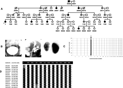
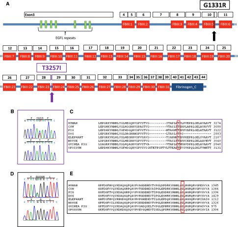
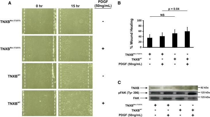
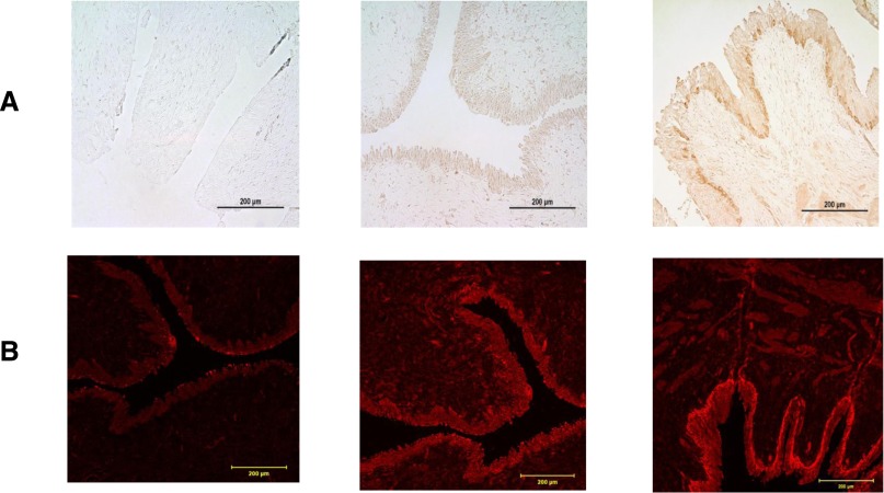
Primary vesicoureteral reflux (VUR) is the most common congenital anomaly of the kidney and the urinary tract, and it is a major risk factor for pyelonephritic scarring and CKD in children. Although twin studies support the heritability of VUR, specific genetic causes remain elusive. We performed a sequential genome-wide linkage study and whole-exome sequencing in a family with hereditary VUR. We obtained a significant multipoint parametric logarithm of odds score of 3.3 on chromosome 6p, and whole-exome sequencing identified a deleterious heterozygous mutation (T3257I) in the gene encoding tenascin XB (TNXB in 6p21.3). This mutation segregated with disease in the affected family as well as with a pathogenic G1331R change in another family. Fibroblast cell lines carrying the T3257I mutation exhibited a reduction in both cell motility and phosphorylated focal adhesion kinase expression, suggesting a defect in the focal adhesions that link the cell cytoplasm to the extracellular matrix. Immunohistochemical studies revealed that the human uroepithelial lining of the ureterovesical junction expresses TNXB, suggesting that TNXB may be important for generating tensile forces that close the ureterovesical junction during voiding. Taken together, these results suggest that mutations in TNXB can cause hereditary VUR.
Figures
Comment in
-
Vesicoureteral reflux: Association of TNXB mutations with vesicoureteral reflux.Nat Rev Nephrol. 2013 Jul;9(7):370. doi: 10.1038/nrneph.2013.90. Epub 2013 May 14. Nat Rev Nephrol. 2013. PMID: 23670083 No abstract available.
References
-
- Arant BS, Jr: Vesicoureteric reflux and renal injury. Am J Kidney Dis 17: 491–511, 1991 - PubMed
-
- North American Pediatric Renal Trials and Collaborative Studies: NAPRTCS 2010 Annual Transplant Report, Boston, MA, NAPRTCS, 2010
-
- Kume T, Deng K, Hogan BL: Murine forkhead/winged helix genes Foxc1 (Mf1) and Foxc2 (Mfh1) are required for the early organogenesis of the kidney and urinary tract. Development 127: 1387–1395, 2000 - PubMed
-
- Ichikawa I, Kuwayama F, Pope JC, 4th, Stephens FD, Miyazaki Y: Paradigm shift from classic anatomic theories to contemporary cell biological views of CAKUT. Kidney Int 61: 889–898, 2002 - PubMed
Publication types
MeSH terms
Substances
Grants and funding
- P30 AG028377/AG/NIA NIH HHS/United States
- RC4 DK090937/DK/NIDDK NIH HHS/United States
- P20 MD006899/MD/NIMHD NIH HHS/United States
- R21 DK096200/DK/NIDDK NIH HHS/United States
- K08 DK082495/DK/NIDDK NIH HHS/United States
- K08DK082495-03/DK/NIDDK NIH HHS/United States
- RC2 NS070342/NS/NINDS NIH HHS/United States
- R56 AI098588/AI/NIAID NIH HHS/United States
- Z01 HG200362/ImNIH/Intramural NIH HHS/United States
- 1R56AI098588-01A1/AI/NIAID NIH HHS/United States
- RC4DK090937/DK/NIDDK NIH HHS/United States
- RC2 MH089915/MH/NIMH NIH HHS/United States
- RC2MH089915/MH/NIMH NIH HHS/United States
- P30AG028377/AG/NIA NIH HHS/United States
- 1RC2NS070342-01/NS/NINDS NIH HHS/United States
LinkOut - more resources
Full Text Sources
Other Literature Sources
Molecular Biology Databases
Miscellaneous
