. 2013 Jul;41(7):1404-13.
doi: 10.1124/dmd.112.050021. Epub 2013 Apr 25.

The effect of breast cancer resistance protein, multidrug resistant protein 1, and organic anion-transporting polypeptide 1B3 on the antitumor efficacy of the lipophilic camptothecin 7-t-butyldimethylsilyl-10-hydroxycamptothecin (AR-67) in vitro

Affiliations

The effect of breast cancer resistance protein, multidrug resistant protein 1, and organic anion-transporting polypeptide 1B3 on the antitumor efficacy of the lipophilic camptothecin 7-t-butyldimethylsilyl-10-hydroxycamptothecin (AR-67) in vitro

Eleftheria Tsakalozou et al. Drug Metab Dispos. 2013 Jul.

Abstract

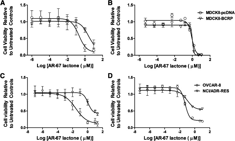
AR-67 (7-t-butyldimethylsilyl-10-hydroxycamptothecin) is a lipophilic camptothecin analog, currently under early stage clinical trials. Transporters are known to have an impact on the disposition of camptothecins and on the response to chemotherapeutics in general due to their expression in tumor tissues. Therefore, we investigated the interplay between the breast cancer resistance protein (BCRP), multidrug resistant protein 1 (MDR1), and organic anion-transporting polypeptide (OATP) 1B1/1B3 transporters and AR-67 and their impact on the toxicity profile of AR-67. Using cell lines expressing the aforementioned transporters, we showed that the lipophilic AR-67 lactone form is a substrate for efflux transporters BCRP and MDR1. Additionally, OATP1B1 and OATP1B3 facilitated the uptake of AR-67 carboxylate in SLCO1B1- and SLCO1B3-transfected cell systems compared with the mock-transfected ones. Notably, both BCRP and MDR1 conferred resistance to AR-67 lactone. Prompted by recent studies showing increased OATP1B3 expression in certain cancer types, we investigated the effect of OATP1B3 expression on cell viability after exposure to AR-67 carboxylate. OATP1B3-expressing cells had increased carboxylate uptake as compared with mock-transfected cells but were not sensitized because the intracellular amount of lactone was 50-fold higher than that of carboxylate and comparable between OATP1B3-expressing and OATP1B3-nonexpressing cells. In conclusion, BCRP- and MDR1-mediated efflux of AR-67 lactone confers resistance to AR-67, but OATP1B3-mediated uptake of the AR-67 carboxylate does not sensitize OATP1B3-expressing tumor cells.


Figures

Fig. 1.
Fig. 1.
pH-dependent interconversion between the lactone and carboxylate form of the camptothecin analog AR-67.
Fig. 2.
Fig. 2.
Effect of BCRP and MDR1 expression on the intracellular AR-67 amounts in MDCKII-pcDNA/BCRP and OVCAR-8/NCI/ADR-RES cells. (A) MDCKII-pcDNA/BCRP and (B) OVCAR-8/NCI/ADR-RES cells were incubated with 0.125, 0.25, 0.5, 0.75 and 1 μM of AR-67 lactone for 5 minutes, and the AR-67 intracellular amount was evaluated. The effect of the dual BCRP/MDR1 inhibitor GF120918 was tested by treating (C) MDCKII-pcDNA/BCRP and (D) OVCAR-8/NCI/ADR-RES cells without (open bars) and with (solid bars) GF120918 (5 µM for 1 hour) before incubating with 1 µM AR-67 lactone for 5 minutes. At the end of the incubation periods, the cells were lysed, and the intracellular AR-67 was quantified using HPLC as mentioned in the Materials and Methods section. Data are represented as mean (n = 3) ± S.D. Statistical analysis was performed using unpaired t test, statistical significance for **P < 0.01; ***P < 0.001.
Fig. 3.
Fig. 3.
Effect of BCRP on the vectorial transport of AR-67 in MDCKII-pcDNA/BCRP cells. Transepithelial transport of 5 µM AR-67 lactone in MDCKII-pcDNA and MDCKII-BCRP cells apical to basolateral (A > B; A and B) and basolateral to apical (B > A; C and D) in the presence (□) and absence (▿) of the inhibitor GF120918 (5 µM). AR-67 was quantified using HPLC as determined in the Materials and Methods section. Data are represented as the mean (n = 3) ± S.D.
Fig. 4.
Fig. 4.
Effect of OATP1B3 and OATP1B1 expression on the intracellular AR-67 amounts in HeLa-pIRES/OATP1B3 and RKO-pIRES/OATP1B1 cells. (A) HeLa-pIRES/OATP1B3 and (B) RKO-pIRES/OATP1B1 cells were incubated with 0.5, 1.0, 1.5, 2.0, and 3.0 µM of AR67 carboxylate for 1 minute to study the dose-dependent uptake of the carboxylate form. The estimated Km values for OATP1B3 and OATP1B1 were 0.32 (0.0–0.8316) μM and 2.58 (0.0–6.253) μM, respectively. They were obtained by using the Michaelis-Menten equation to fit the transporter-mediated uptake of AR-67, as described in the Materials and Methods section, and are reported as the mean (95% confidence interval). (C) HeLa-pIRES/OATP1B3 and (D) RKO-pIRES/OATP1B1 cells were incubated with 1 μM of AR-67 carboxylate for 5 and 10 minutes, respectively (open bars). The inhibitory effect of BSP (50 μM) was studied by pretreating for 10 minutes before exposure to AR-67 carboxylate as described previously for all cell lines (solid bars). Intracellular AR-67 was quantified using HPLC, as determined in the Materials and Methods section. Data are represented as the mean (n = 3) ± S.D. Statistical analysis was performed using unpaired t test, statistical significance for **P < 0.01; ***P < 0.001.
Fig. 5.
Fig. 5.
BCRP and MDR1 decrease the cytotoxicity of AR-67 lactone. (A) After 72-hour drug incubation, IC50 values were estimated in MDCKII-pcDNA (0.21 μM, [0.11–0.40]) and MDCKII-BCRP (2.37 μM, [1.41–4.00]; P < 0.01, unpaired Student’s t test). (B) After pretreatment (30 minutes) and in the presence of GF120918 and AR-67 lactone (72 hours), IC50 values were obtained in MDCKII-pcDNA (0.88 μM, [0.72–1.09]) and MDCKII-BCRP (1.02 μM, [0.79–1.32]). (C) Similarly, IC50 values were estimated in OVCAR-8 (0.027 μM, [0.018–0.043]) and NCI/ADR-RES (1.16 μM, [0.79–1.70]; P < 0.0001, unpaired Student’s t test). (D) After pretreatment (30 minutes) and in the presence of GF120918 and AR-67 lactone (72 hours), IC50 values were obtained in OVCAR-8 (0.053 μM, [0.044–0.064]) and NCI/ADR-RES cells (0.095 μM, [0.044–0.21]). The AR-67 lactone doses ranged from 10−7 to 21 µM. IC50 parameters and best-fit lines were estimated by nonlinear regression analysis. Data are plotted as the mean ± S.D. (n = 3), and IC50 values (μM) are reported as the mean (95% confidence interval).
Fig. 6.
Fig. 6.
Time-dependent cytotoxic effect of AR-67 carboxylate on HeLa-pIRES and HeLa-OATP1B3 cell lines. (A–D) HeLa-pIRES and HeLa-OATP1B3 cell lines were exposed to AR-67 carboxylate for 30 minutes (A), 1 hour (B), 3 hours (C), and 48 hours (D), as described in the Materials and Methods section. The AR-67 carboxylate doses ranged from 10−3 to 210 µM. (E) Summary of the cytotoxicity of AR-67 carboxylate on HeLa-pIRES and HeLa-OATP1B3 cells studied under the experimental conditions described in panels A–D. IC50 values to reflect the effect of AR-67 on cell viability were used. Cell viability was assessed at the end of the treatment as described in the Materials and Methods section. Nonlinear regression analysis was performed to model the data (solid line). Data points on the graphs are reported as the mean (n = 3) ± S.D., and IC50 values (μM) are reported as the mean (95% confidence interval).
Fig. 7.
Fig. 7.
Stability of AR-67 in cell culture media. The time course of AR-67 interconversion was studied in cell culture media (pH 7.4, 37°C, 5% CO2) after the addition of 1 μM of AR-67 carboxylate (A) or 1 μM of AR-67 lactone (B). Data are represented as the percentage ratio concentration of carboxylate or lactone over total drug.
Fig. 8.
Fig. 8.
Time course of AR-67 carboxylate and lactone intracellular amount in HeLa-pIRES and HeLa-OATP1B3 cells after incubation with AR-67 carboxylate or lactone. HeLa-pIRES and HeLa-OATP1B3 cells were incubated with 1 μM of the AR-67 forms, carboxylate (A) and lactone (B). AR-67 forms intracellularly were quantified by HPLC as described in the Materials and Methods section. Data are represented as the mean (n = 3) ± S.D.
Fig. 9.
Fig. 9.
Effect of AR-67 carboxylate and lactone on HeLa-pIRES and HeLa-OATP1B3 cell lines. (A) HeLa-pIRES and HeLa-OATP1B3 cells were treated with AR-67 carboxylate for 5 minutes, cells were washed, drug-free medium was added into the wells, and the cells were allowed to grow for 48 hours before assessing cell viability. The estimated IC50 values were 40.32 μM (16.36–99.33) and 13.08 μM (8.29–20.62) for HeLa-pIRES and HeLa-OATP1B3 cells, respectively. (B) HeLa-pIRES and HeLa-OATP1B3 cells were treated with AR-67 lactone for 5 minutes, cells were washed, and drug-free medium was added into the wells. The estimated IC50 values were 1.08 μM (0.20–5.70) and 0.23 μM (0.028–1.93) for HeLa-pIRES and HeLa-OATP1B3 cells, respectively. Data analysis to obtain IC50 values was performed using nonlinear regression (solid line). Data are presented as the mean (n = 3) ± S.D., and IC50 values (μM) are reported as the mean (95% confidence interval).

References

    1. Abe T, Kakyo M, Tokui T, Nakagomi R, Nishio T, Nakai D, Nomura H, Unno M, Suzuki M, Naitoh T, et al. (1999) Identification of a novel gene family encoding human liver-specific organic anion transporter LST-1. J Biol Chem 274:17159–17163 - PubMed
    1. Abe T, Unno M, Onogawa T, Tokui T, Kondo TN, Nakagomi R, Adachi H, Fujiwara K, Okabe M, Suzuki T, et al. (2001) LST-2, a human liver-specific organic anion transporter, determines methotrexate sensitivity in gastrointestinal cancers. Gastroenterology 120:1689–1699 - PubMed
    1. Adane ED, Liu Z, Xiang TX, Anderson BD, Leggas M. (2010) Factors affecting the in vivo lactone stability and systemic clearance of the lipophilic camptothecin analogue AR-67. Pharm Res 27:1416–1425 - PubMed
    1. Arnold SM, Rinehart JJ, Tsakalozou E, Eckardt JR, Fields SZ, Shelton BJ, DeSimone PA, Kee BK, Moscow JA, Leggas M. (2010) A phase I study of 7-t-butyldimethylsilyl-10-hydroxycamptothecin in adult patients with refractory or metastatic solid malignancies. Clin Cancer Res 16:673–680 - PMC - PubMed
    1. Bom D, Curran DP, Kruszewski S, Zimmer SG, Thompson Strode J, Kohlhagen G, Du W, Chavan AJ, Fraley KA, Bingcang AL, et al. (2000) The novel silatecan 7-tert-butyldimethylsilyl-10-hydroxycamptothecin displays high lipophilicity, improved human blood stability, and potent anticancer activity. J Med Chem 43:3970–3980 - PubMed

Publication types

MeSH terms

Substances