p63-expressing cells are the stem cells of developing prostate, bladder, and colorectal epithelia
- PMID: 23620512
- PMCID: PMC3657776
- DOI: 10.1073/pnas.1221216110
p63-expressing cells are the stem cells of developing prostate, bladder, and colorectal epithelia
Abstract
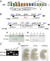
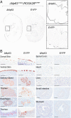
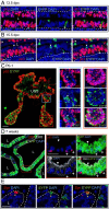
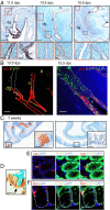
The tumor protein p63 (p63), and more specifically the NH2-terminal truncated (ΔN) p63 isoform, is a marker of basal epithelial cells and is required for normal development of several epithelial tissues, including the bladder and prostate glands. Although p63-expressing cells are proposed to be the stem cells of the developing prostate epithelium and bladder urothelium, cell lineages in these endoderm-derived epithelia remain highly controversial, and rigorous lineage tracing studies are warranted. Here, we generated knock-in mice expressing Cre recombinase (Cre) under the control of the endogenous ΔNp63 promoter. Heterozygote ΔNp63(+/Cre) mice were phenotypically normal and fertile. Cre-mediated recombination in ΔNp63(+/Cre);ROSA26(EYFP) reporter mice faithfully recapitulated the pattern of ΔNp63 expression and were useful for genetic lineage tracing of ΔNp63-expressing cells of the caudal endoderm in vivo. We found that ΔNp63-positive cells of the urogenital sinus generated all epithelial lineages of the prostate and bladder, indicating that these cells represent the stem/progenitor cells of those epithelia during development. We also observed ΔNp63 expression in caudal gut endoderm and the contribution of ΔNp63-positive cells to the stem/progenitor compartment of adult colorectal epithelium. Because p63 is a master regulator of stratified epithelial development, this finding provides a unique developmental insight into the cell of origin of squamous cell metaplasia and squamous cell carcinoma of the colon.
Keywords: genitourinary tract; hindgut; large intestine.
Conflict of interest statement
The authors declare no conflict of interest.
Figures


Similar articles
-
Expression of p53-related protein p63 in the gastrointestinal tract and in esophageal metaplastic and neoplastic disorders.Hum Pathol. 2001 Nov;32(11):1157-65. doi: 10.1053/hupa.2001.28951. Hum Pathol. 2001. PMID: 11727253
-
Differential expression of p63 isoforms in normal tissues and neoplastic cells.J Pathol. 2002 Dec;198(4):417-27. doi: 10.1002/path.1231. J Pathol. 2002. PMID: 12434410
-
ΔNp63 knockout mice reveal its indispensable role as a master regulator of epithelial development and differentiation.Development. 2012 Feb;139(4):772-82. doi: 10.1242/dev.071191. Development. 2012. PMID: 22274697 Free PMC article.
-
Taking advantage of basic research: p63 is a reliable myoepithelial and stem cell marker.Adv Anat Pathol. 2002 Sep;9(5):280-9. doi: 10.1097/00125480-200209000-00002. Adv Anat Pathol. 2002. PMID: 12195217 Review.
-
Dynamic life of a skin keratinocyte: an intimate tryst with the master regulator p63.Indian J Exp Biol. 2011 Oct;49(10):721-31. Indian J Exp Biol. 2011. PMID: 22013738 Review.
Cited by
-
ΔN63 suppresses the ability of pregnancy-identified mammary epithelial cells (PIMECs) to drive HER2-positive breast cancer.Cell Death Dis. 2021 May 22;12(6):525. doi: 10.1038/s41419-021-03795-5. Cell Death Dis. 2021. PMID: 34023861 Free PMC article.
-
Inducible knockout of ∆Np63 alters cell polarity and metabolism during pubertal mammary gland development.FEBS Lett. 2020 Mar;594(6):973-985. doi: 10.1002/1873-3468.13703. Epub 2019 Dec 17. FEBS Lett. 2020. PMID: 31794060 Free PMC article.
-
The DeltaN p63 Promotes EMT and Metastasis in Bladder Cancer by the PTEN/AKT Signalling Pathway.Evid Based Complement Alternat Med. 2022 Apr 12;2022:9566055. doi: 10.1155/2022/9566055. eCollection 2022. Evid Based Complement Alternat Med. 2022. PMID: 35463095 Free PMC article.
-
Prostate stem cells in the development of benign prostate hyperplasia and prostate cancer: emerging role and concepts.Biomed Res Int. 2013;2013:107954. doi: 10.1155/2013/107954. Epub 2013 Jul 8. Biomed Res Int. 2013. PMID: 23936768 Free PMC article. Review.
-
Retinoid signaling in progenitors controls specification and regeneration of the urothelium.Dev Cell. 2013 Sep 16;26(5):469-482. doi: 10.1016/j.devcel.2013.07.017. Epub 2013 Aug 29. Dev Cell. 2013. PMID: 23993789 Free PMC article.
References
-
- Marker PC, Donjacour AA, Dahiya R, Cunha GR. Hormonal, cellular, and molecular control of prostatic development. Dev Biol. 2003;253(2):165–174. - PubMed
-
- Goto K, et al. Proximal prostatic stem cells are programmed to regenerate a proximal-distal ductal axis. Stem Cells. 2006;24(8):1859–1868. - PubMed
Publication types
MeSH terms
Substances
Grants and funding
LinkOut - more resources
Full Text Sources
Other Literature Sources
Medical
Molecular Biology Databases
Research Materials

