. 2013 May 7;110(19):7916-21.
doi: 10.1073/pnas.1300677110. Epub 2013 Apr 25.

Modulation of the endoplasmic reticulum-mitochondria interface in Alzheimer's disease and related models

Affiliations

Modulation of the endoplasmic reticulum-mitochondria interface in Alzheimer's disease and related models

Louise Hedskog et al. Proc Natl Acad Sci U S A. .

Abstract

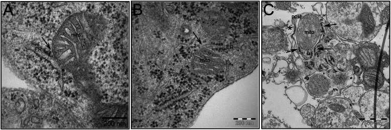
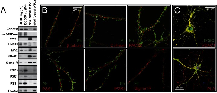
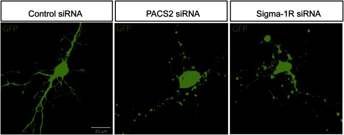
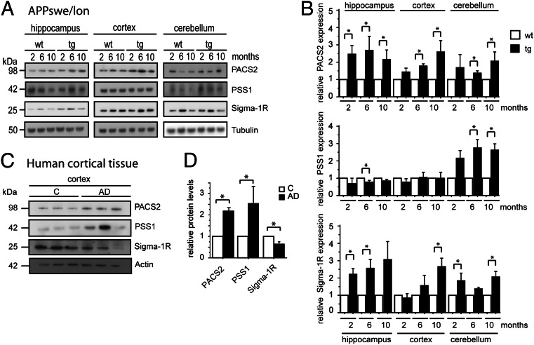
It is well-established that subcompartments of endoplasmic reticulum (ER) are in physical contact with the mitochondria. These lipid raft-like regions of ER are referred to as mitochondria-associated ER membranes (MAMs), and they play an important role in, for example, lipid synthesis, calcium homeostasis, and apoptotic signaling. Perturbation of MAM function has previously been suggested in Alzheimer's disease (AD) as shown in fibroblasts from AD patients and a neuroblastoma cell line containing familial presenilin-2 AD mutation. The effect of AD pathogenesis on the ER-mitochondria interplay in the brain has so far remained unknown. Here, we studied ER-mitochondria contacts in human AD brain and related AD mouse and neuronal cell models. We found uniform distribution of MAM in neurons. Phosphofurin acidic cluster sorting protein-2 and σ1 receptor, two MAM-associated proteins, were shown to be essential for neuronal survival, because siRNA knockdown resulted in degeneration. Up-regulated MAM-associated proteins were found in the AD brain and amyloid precursor protein (APP)Swe/Lon mouse model, in which up-regulation was observed before the appearance of plaques. By studying an ER-mitochondria bridging complex, inositol-1,4,5-triphosphate receptor-voltage-dependent anion channel, we revealed that nanomolar concentrations of amyloid β-peptide increased inositol-1,4,5-triphosphate receptor and voltage-dependent anion channel protein expression and elevated the number of ER-mitochondria contact points and mitochondrial calcium concentrations. Our data suggest an important role of ER-mitochondria contacts and cross-talk in AD pathology.

Keywords: AD mouse models; hippocampal neurons; human cortical brain tissue.


Conflict of interest statement

The authors declare no conflict of interest.

Figures

Fig. 1.
Fig. 1.
EM pictures of the ER–mitochondria contact point in mouse brain tissue and hippocampal neurons. MAM structures are indicated by arrows in (A) embryonic day 16 mouse hippocampal tissue, (B) primary mouse hippocampal neurons, and (C) synaptosomes prepared from adult mouse brain. The synaptic cleft and postsynapse are indicated by * and post, respectively. (Scale bars: A and B, 200 nm; C, 500 nm.)
Fig. 2.
Fig. 2.
Analysis of MAM-associated proteins. Western blot analysis of subcellularly fractionated mouse brain and ICC staining of hippocampal neurons. (A) Mouse brain homogenate was fractionated into 20,000 × g (P3) containing, for example, plasma membrane, 100,000 × g (P4) containing, for example, ER, mito (percoll of P2), and MAM (percoll of P2); 25 µg protein from each fraction were loaded onto the gel. Markers for subcellular fractions were calnexin (ER), Na/K-ATPase (plasma membrane), COX1 (inner mitochondrial membrane), and GM130 (Golgi). (B) Confocal microscopy of neurons stained with phalloidin (Ph; spines), β-tubulin (neuronal marker), calnexin (ER), and MAM-associated proteins (PACS-2, PSS1, IP3R3, and σ1R). 100× objective. (C, Upper) Distribution of IP3R3 (green; ER side) and VDAC1 (red; mitochondrial side). (C, Lower) PLA signals (reds dots) show close proximity between IP3R3 and VDAC1, indicating interactions between ER and mitochondrial membranes.
Fig. 3.
Fig. 3.
MAM-associated protein depletion causes degeneration of hippocampal neurons. Hippocampal neurons were transfected with AllStar negative control siRNA or siRNA to PACS-2 or σ1R (30 nM) together with pEGFP-N1 vector (0.4 ng/µL). Live cell images taken 16 h after transfection.
Fig. 4.
Fig. 4.
MAM-associated protein expression is altered in AD and AD mouse model. Western blot quantification of expression of MAM-associated proteins in (A and B) the APPSwe/Lon mouse brain (2, 6, and 10 mo old) and (C and D) the human cortical postmortem brain tissue. Data were compared using Mann–Whitney u test (APPSwe/Lon: mean ± SD, n = 4; human cortical brain tissue: mean ± SEM, n = 3; *P < 0.05). Data were normalized to (B) tubulin or (D) actin.
Fig. 5.
Fig. 5.
Aβ exposure elevates the expression of MAM-associated proteins and increases ER–mitochondrial contacts in hippocampal neurons. (A) Western blot of APP expression in CHO7PA2 cells and CHOwt cells. (B) Aβ levels in the CM at two concentrations: 2.5 (CM) and 4.5 nM (CM*) Aβ40 + Aβ42. (C) Confocal images acquired using the same experimental settings show VDAC1 (red) and IP3R3 (green) fluorescence intensity in untreated and CM*-treated (8 h) hippocampal neurons. (D) Quantification of fluorescence intensity of VDAC1 and IP3R3 after exposure to CM and CM* for 8 and 48 h. Data were compared using ANOVA followed by Games/Howell posthoc test (mean ± SD, n = 4; *P < 0.05). (E) For quantification of VDAC1 and IP3R3 interaction, we performed PLA on 8-h CM*-treated hippocampal neurons. The fluorescent spots (PLA signals) obtained using Duolink detection fluorophore Far-red ex644 were analyzed using Duolink Imaging Tool software. Data were compared using Mann–Whitney u test (mean ± SD, n = 3; *P < 0.05).
Fig. 6.
Fig. 6.
Aβ exposure increases the Ca2+ shuttling from ER to mitochondria. Cytosolic and mitochondrial [Ca2+] measured in SH-SY5Y cells. (A) The σ1R agonist SA4503 does not affect cyt-Δ [Ca2+] or mit-Δ [Ca2+]. (B) Representative figure of Ca2+ peaks after treatment with SA4503 and CM* alone or combined. (C) Nanomolar concentrations of Aβ ± SA4503 do not increase cyt-Δ [Ca2+]. (D) Nanomolar concentrations of CM* + SA4503 increase ER to mitochondria Ca2+ transfer. (E) Treatment with TUN ± SA4503 does not increase cyt-Δ [Ca2+]. (F) Treatment of TUN combined with SA4503 increased ER to mitochondria Ca2+ transfer. (G) CM* increased mitochondrial Ca2+ uptake from cytosol (here exchanged with buffer containing fixed [Ca2+]). Data were compared using ANOVA and Games/Howell posthoc test (mean ± SEM, n > 4; *P < 0.05). Mock, DMSO.

References

    1. Ankarcrona M, Mangialasche F, Winblad B. Rethinking Alzheimer’s disease therapy: Are mitochondria the key? J Alzheimers Dis. 2010;20(Suppl 2):S579–S590. - PubMed
    1. Area-Gomez E, et al. Upregulated function of mitochondria-associated ER membranes in Alzheimer disease. EMBO J. 2012;31(21):4106–4123. - PMC - PubMed
    1. Hayashi T, Rizzuto R, Hajnoczky G, Su TP. MAM: More than just a housekeeper. Trends Cell Biol. 2009;19(2):81–88. - PMC - PubMed
    1. Rusiñol AE, Cui Z, Chen MH, Vance JE. A unique mitochondria-associated membrane fraction from rat liver has a high capacity for lipid synthesis and contains pre-Golgi secretory proteins including nascent lipoproteins. J Biol Chem. 1994;269(44):27494–27502. - PubMed
    1. Vance JE. Molecular and cell biology of phosphatidylserine and phosphatidylethanolamine metabolism. Prog Nucleic Acid Res Mol Biol. 2003;75(2003):69–111. - PubMed

Publication types

MeSH terms