. 2013 Jun 7;288(23):17008-17018.
doi: 10.1074/jbc.M113.468926. Epub 2013 Apr 25.

Substrate-modulated cytochrome P450 17A1 and cytochrome b5 interactions revealed by NMR

Affiliations

Substrate-modulated cytochrome P450 17A1 and cytochrome b5 interactions revealed by NMR

D Fernando Estrada et al. J Biol Chem. .

Abstract

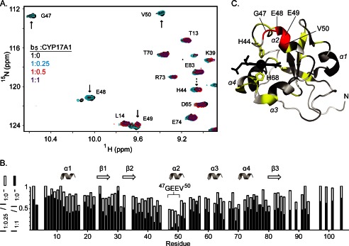
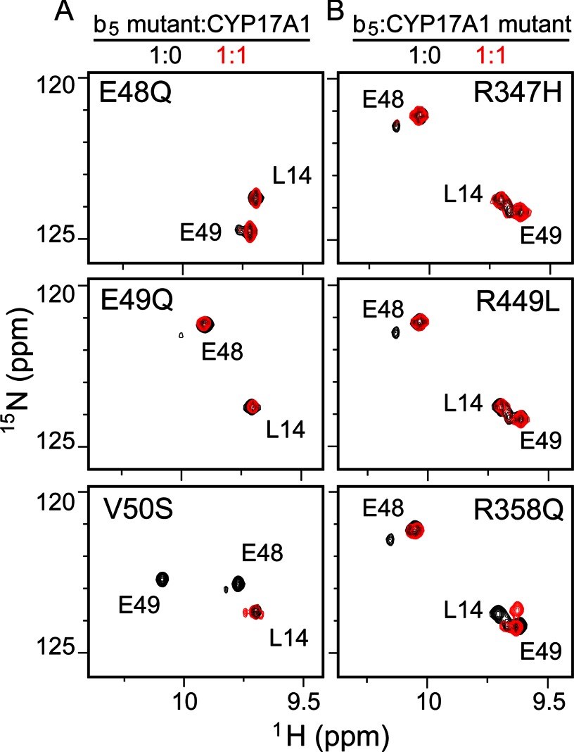
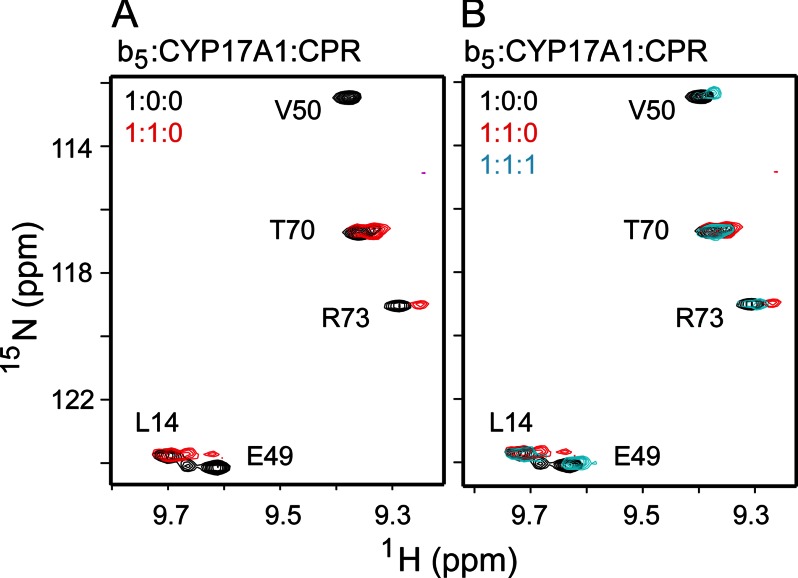
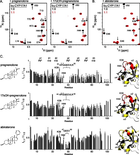
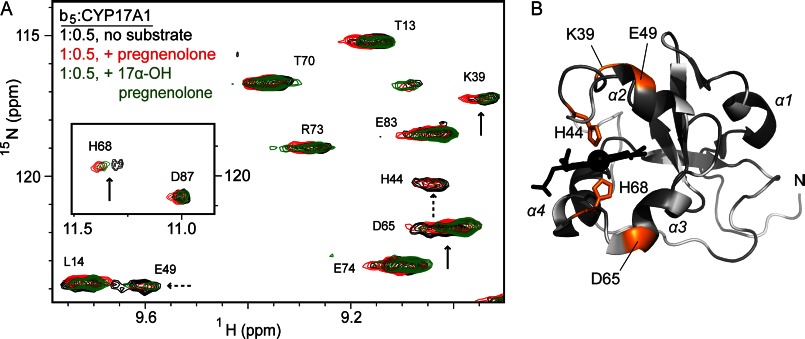
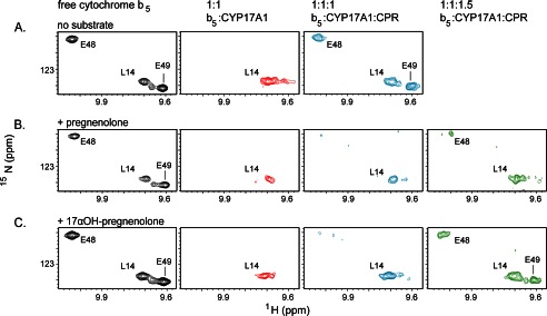
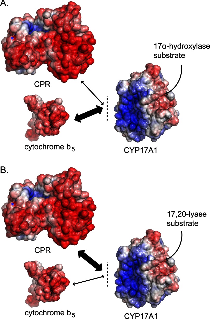
The membrane heme protein cytochrome b5 (b5) can enhance, inhibit, or have no effect on cytochrome P450 (P450) catalysis, depending on the specific P450, substrate, and reaction conditions, but the structural basis remains unclear. Here the interactions between the soluble domain of microsomal b5 and the catalytic domain of the bifunctional steroidogenic cytochrome P450 17A1 (CYP17A1) were investigated. CYP17A1 performs both steroid hydroxylation, which is unaffected by b5, and an androgen-forming lyase reaction that is facilitated 10-fold by b5. NMR chemical shift mapping of b5 titrations with CYP17A1 indicates that the interaction occurs in an intermediate exchange regime and identifies charged surface residues involved in the protein/protein interface. The role of these residues is confirmed by disruption of the complex upon mutagenesis of either the anionic b5 residues (Glu-48 or Glu-49) or the corresponding cationic CYP17A1 residues (Arg-347, Arg-358, or Arg-449). Cytochrome b5 binding to CYP17A1 is also mutually exclusive with binding of NADPH-cytochrome P450 reductase. To probe the differential effects of b5 on the two CYP17A1-mediated reactions and, thus, communication between the superficial b5 binding site and the buried CYP17A1 active site, CYP17A1/b5 complex formation was characterized with either hydroxylase or lyase substrates bound to CYP17A1. Significantly, the CYP17A1/b5 interaction is stronger when the hydroxylase substrate pregnenolone is present in the CYP17A1 active site than when the lyase substrate 17α-hydroxypregnenolone is in the active site. These findings form the basis for a clearer understanding of this important interaction by directly measuring the reversible binding of the two proteins, providing evidence of communication between the CYP17A1 active site and the superficial proximal b5 binding site.

Keywords: Cytochrome P450 17A1; Cytochrome b5; Enzyme Mechanisms; NMR; Protein Conformation; Protein-Protein Interactions; Steroidogenesis.


Figures

FIGURE 1.
FIGURE 1.
Chemical shift mapping of cytochrome b5/CYP17A1 interaction shows binding on an intermediate exchange time scale. A, HSQC spectra of 15N-b5 titrated with unlabeled CYP17A1 show a combination of chemical shift perturbations and line broadening. Residues indicated by solid arrows showed substantial line broadening so that a signal was no longer detectible or very weak at a half-molar equivalent of CYP17A1, whereas small shifts were observed in other residues (dashed arrows). B, peak intensity for two titration points (b5:CYP17A1 1:0.25 in open bars and 1:1 in black bars) plotted as a fraction of the free b5 intensity (b5:CYP17A1 1:0 molar ratio) revealed consistent and disproportionate line broadening for residues 47GEEV50 throughout the titration. C, these broadened residues (red) occur in the b5 α2 helix and its preceding loop. Other residues with small shifts (yellow) include His-44 and His-68 on both sides of the b5 heme, suggesting a change in the heme position. Residues shown in gray have little or no perturbation, whereas those shown in white were not evaluated because of either spectral overlap or the presence of a proline.
FIGURE 2.
FIGURE 2.
Conserved anionic (b5) and cationic (CYP17A1) residues are required to form the b5-CYP17A1 complex. Shown are 2D 15N-HSQC spectra of b5 (1:0, black) overlaid with the same samples containing an equal molar equivalent (1:1, red) of CYP17A1. A, effects of b5 mutation. Broadening of Glu-48 or Glu-49 was not observed in the b5 E48Q and E49Q mutants (top and center panels, respectively), suggesting that the b5/CYP17A1 complex was not formed. However, the b5 V50S mutant did show broadening of both resonances (bottom panel), suggesting b5-CYP17A1 complex formation similar to wild-type b5. B, effects of CYP17A1 mutation. None of the CYP17A1 R347H, R358Q, and R449L mutant proteins induced line broadening in either Glu-48 or Glu-49, suggesting that the b5-CYP17A1 complex was not formed.
FIGURE 3.
FIGURE 3.
Addition of P450 reductase disrupts the b5-CYP17A1 complex. Representative segments of the overlaid 2D 15N-HSQC spectra demonstrated that b5/CYP17A1 binding (shown in the absence of CPR in A in red) can be disrupted by adding an equal molar amount of full-length rat CPR, as indicated by the return of the liberated b5 spectrum (B, blue).
FIGURE 4.
FIGURE 4.
Alterations in b5-CYP17A1 complex formation in the presence of CYP17A1 ligands including the 17α-hydroxylase substrate pregnenolone, the 17,20-lyase substrate 17α-hydroxypregnenolone, and the heme-coordinating inhibitor abiraterone. A, representative HSQC spectra of 15N-b5 alone (1:0) and in the presence of an equimolar concentration of CYP17A1 (b5/CYP17A1 1:1) were collected in the presence of each substrate, showing that in addition to broadening of 47GEEV50 observed during b5 binding to unliganded CYP17A1 (solid arrows), enhanced line broadening also occurred for specific residues near the binding interface (dashed arrows). B, the steroidal inhibitor abiraterone coordinates the CYP17A1 heme iron via its pyridine ring, resulting in a low-spin system that is less strongly paramagnetic, allowing distinction of paramagnetic effects on residues like His-44, Lys-39, and Glu-49 (dashed arrows) from intermediate exchange effects on Glu-48 and Val-50 because of complex formation (solid arrows). C, plots of the relative intensities for all b5 resonances in the ligand bound versus free state illustrate substrate-induced reductions in intensity because of line broadening in regions adjoining the 47GEEV50 sequence for substrates but not for the inhibitor abiraterone. Residues marked with an asterisk are located near the binding site but were either not assigned or consist of a proline. For each liganded condition, residues line-broadened below 15% of the original signal are mapped onto the structure in red in the right panel.
FIGURE 5.
FIGURE 5.
CYP17A1 substrate differentially affects CYP17A1/b5 interaction. A, representative spectra of b5 in the presence of a half-molar equivalent of CYP17A1:substrate. At a reduced substrate-bound CYP17A1 concentration, b5 line broadening is less severe. As a result, minor differences can be observed in the binding to unliganded (black), pregnenolone bound (red), and 17α-hydroxypregnenolone bound (green) CYP17A1. In particular, Lys-39, Asp-65, and His-68 are perturbed differently for each liganded state (black arrows), whereas His-44 and Glu-49 (dashed arrows) are both perturbed as well as line-broadened to a different extent. B, the differentially affected residues (orange) map onto both sides of b5, suggestive of a substrate-influenced change in the b5 heme.
FIGURE 6.
FIGURE 6.
CYP17A1 substrate differentially affects the ability of CPR to disrupt binding to b5. Samples containing 15N-b5 and CYP17A1 in 1:1 molar ratios were titrated with CPR, and HSQC spectra were reacquired. A, as demonstrated, in the absence of CYP17A1 substrate, an equal molar amount of CPR (1:1:1) was sufficient to observe the complete return of resonances for Glu-48 and Glu-49, indicating the liberation of free b5. B, when pregnenolone is bound to CYP17A1, a greater molar equivalent of CPR was required to liberate free b5, as indicated by a weak Glu-48 and Glu-49 signal even with 1.5 molar equivalents of CPR. C, when 17α-hydroxypregnenolone is the CYP17A1 ligand, more CPR is required to liberate b5 than in the substrate-free situation, as indicated by weak signals in the 1:1:1 condition, but the free b5 signal fully reappears upon increasing CPR to the 1.5 molar equivalent (1:1:1.5).
FIGURE 7.
FIGURE 7.
Model describing the second-order effects of 17α-hydroxylase and 17,20-lyase substrates on modulation of the b5-CYP17A1 and CPR-CYP17A1 complexes. A, the current data support a scheme in which, when in the presence of pregnenolone, the b5-CYP17A1 electrostatically driven association is enhanced relative to the same interaction in the absence of substrate, thus resulting in decreased access to electron transfer via CPR. B, the scales of this balance tip in the presence of 17α-hydroxypregnenolone, in which a weaker b5:CYP17A1 association (relative to pregnenolone) results in enhanced electron transfer via CPR.

Similar articles

Cited by

References

    1. Ortiz de Montellano P. R. (2005) Cytochrome P450. Structure, Mechanism, and Biochemistry, 3rd ed., pp. 133–134 and 448–450, Kluwer Academic/Plenum Publishers, New York
    1. Schenkman J. B., Jansson I. (2003) The many roles of cytochrome b5. Pharmacol. Ther. 97, 139–152 - PubMed
    1. Im S. C., Waskell L. (2011) The interaction of microsomal cytochrome P450 2B4 with its redox partners, cytochrome P450 reductase and cytochrome b(5). Arch. Biochem. Biophys. 507, 144–153 - PMC - PubMed
    1. Zhang H., Im S. C., Waskell L. (2007) Cytochrome b5 increases the rate of product formation by cytochrome P450 2B4 and competes with cytochrome P450 reductase for a binding site on cytochrome P450 2B4. J. Biol. Chem. 282, 29766–29776 - PubMed
    1. Akhtar M. K., Kelly S. L., Kaderbhai M. A. (2005) Cytochrome b(5) modulation of 17α-hydroxylase and 17–20 lyase (CYP17) activities in steroidogenesis. J. Endocrinol. 187, 267–274 - PubMed

Publication types

MeSH terms

LinkOut - more resources