

Role of (18)F-FDG PET-CT in head and neck squamous cell carcinoma
- PMID: 23620633
- PMCID: PMC3631810
Role of (18)F-FDG PET-CT in head and neck squamous cell carcinoma
Abstract
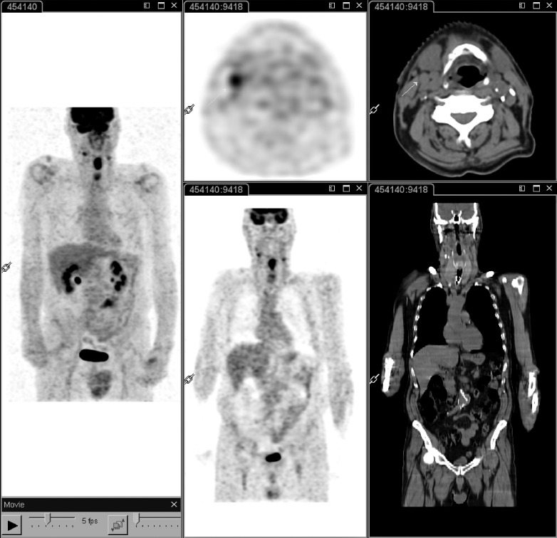
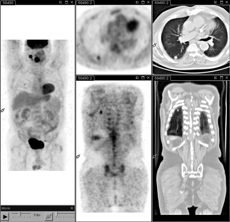
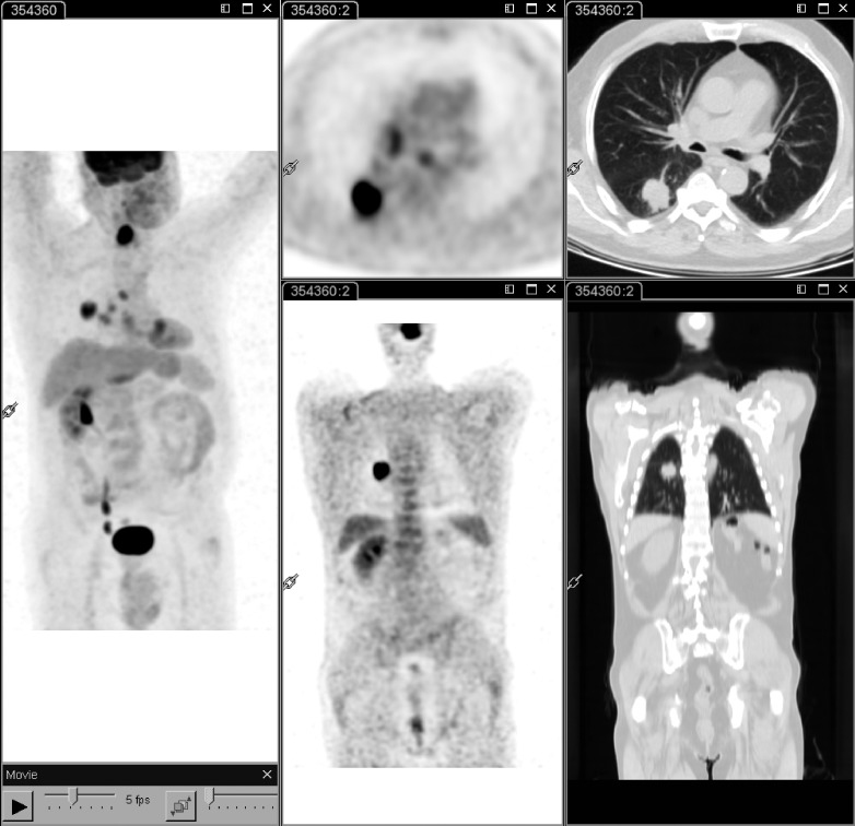
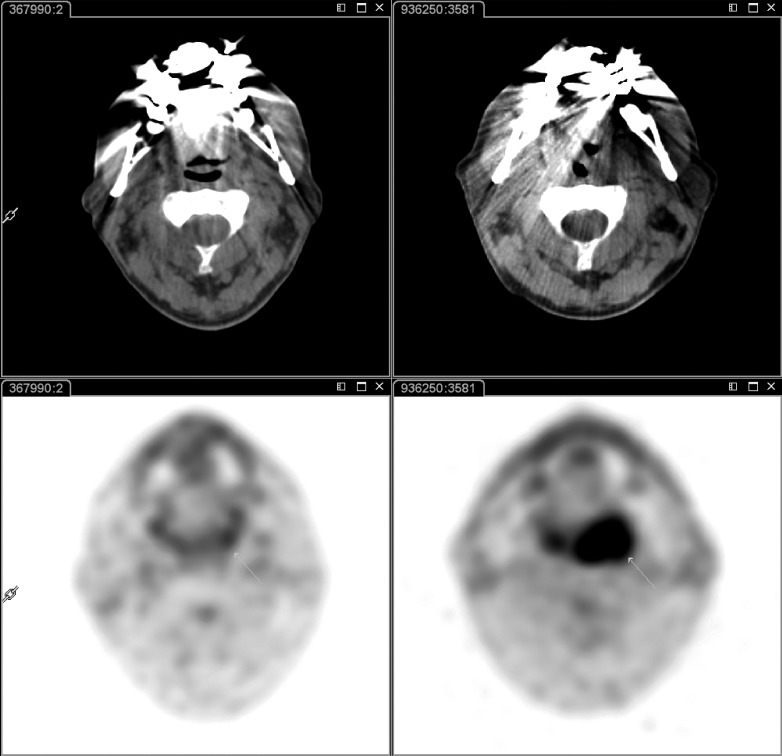
The role of PET-CT imaging in head and neck squamous cell carcinoma during pre-treatment staging, radiotherapy planning, treatment response assessment and post-therapy follow-up is reviewed with focus on current evidence, controversial issues and future clinical applications. In staging, the role of (18)F-FDG PET-CT is well recognized for detecting cervical nodal involvement as well as for exclusion of distant metastases and synchronous primary tumours. In the evaluation of treatment response, the high negative predictive value of (18)F-FDG PET-CT performed at least 8 weeks from the end of radio-chemotherapy allows prevention of unnecessary diagnostic invasive procedures and neck dissection in many patients, with a significant impact on clinical outcome. On the other hand, in this setting, the low positive predictive value due to possible post-radiation inflammation findings requires special care before making a clinical decision. Controversial data are currently available on the role of PET imaging during the course of radio-chemotherapy. The prognostic role of (18)F-FDG PET-CT imaging in head and neck squamous cell carcinoma is recently emerging, in addition to the utility of this technique in evaluation of the tumour volume for planning radiation therapy. Additionally, new PET radiopharmaceuticals could provide considerable information on specific tumour characteristics, thus overcoming the limitations of (18)F-FDG.
In questa review è analizzato il ruolo della PET-CT nei carcinomi squamosi del distretto testa-collo in fase di stadiazione, nella pianificazione del trattamento radiante, nella valutazione della risposta al trattamento radio-chemioterapico e nel follow-up, tenendo conto dei dati attualmente disponibili, delle questioni controverse e delle future applicazioni cliniche. In fase di stadiazione, è ampiamente riconosciuto il ruolo della PET-CT con 18F-FDG nella valutazione del coinvolgimento linfonodale, nonché nella esclusione della presenza di metastasi a distanza e di tumori primitivi sincroni. Nella valutazione della risposta al trattamento, l'elevato valore predittivo negativo della 18F-FDG PET-CT, effettuata almeno 8 settimane dopo la fine del trattamento radio-chemioterapico, consente di evitare in molti pazienti inutili procedure diagnostiche invasive nonché la dissezione del collo, con conseguente significativo impatto clinico. D'altra parte, in questa fase il basso valore predittivo positivo della metodica, causato dai possibili falsi positivi secondari alla concomitante flogosi post-attinica, deve essere tenuto in particolare considerazione prima di prendere una decisione clinica. Dati controversi sono attualmente disponibili sul ruolo dell'imaging PET durante il trattamento radio-chemioterapico. Negli ultimi anni, è emerso il ruolo prognostico della 18F-FDG PET-CT nei carcinomi a cellule squamose del distretto testa-collo, così come l'utilità di questa tecnica nella valutazione del volume del tumore per la pianificazione del trattamento radiante. Inoltre, nuove prospettive provengono dall'impiego dei nuovi radiofarmaci PET che potrebbero fornire notevoli informazioni su caratteristiche biologiche specifiche del tumore, superando di conseguenza i noti limiti del 18F-FDG.
Keywords: 18F-FDG; Carcinoma; Head and neck tumours; PET-CT; Squamous cell.
Figures
References
-
- Oyen WJ, Marres HA, Kaanders JH. Progress in nuclear medicine procedures in head and neck oncology. Q J Nucl Med Mol Imaging. 2011;55:485–486. - PubMed
-
- Corvò R. Evidence-based radiation oncology in head and neck squamous cell carcinoma. Radiother Oncol. 2007;85:156–170. - PubMed
-
- Schwarz E, Hurlimann S, Soyka JD, et al. FDG-positive Warthin's tumors in cervical lymph nodes mimicking metastases in tongue cancer staging with PET/CT. Otolaryngol Head Neck Surg. 2009;140:134–135. - PubMed
-
- Kubota R, Yamada S, Kubota K, et al. Intratumoral distribution of fluorine-18-fluorodeoxyglucose in vivo: high accumulation in macrophages and granulation tissues studied by microautoradiography. J Nucl Med. 1992;33:1972–1980. - PubMed
-
- Hentschel M, Appold S, Schreiber A, et al. Serial FDG-PET on patients with head and neck cancer: implications for radiation therapy. Int J Radiat Biol. 2009;85:796–804. - PubMed
Publication types
MeSH terms
Substances
LinkOut - more resources
Full Text Sources
Medical