

Clinical significance of metallothioneins in cell therapy and nanomedicine
- PMID: 23620664
- PMCID: PMC3633583
- DOI: 10.2147/IJN.S42019
Clinical significance of metallothioneins in cell therapy and nanomedicine
Abstract
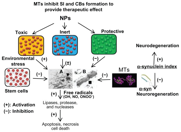
Mammalian metallothioneins (MTs) are low molecular weight (6-7 kDa) cysteine-rich proteins that are specifically induced by metal nanoparticles (NPs). MT induction in cell therapy may provide better protection by serving as antioxidant, anti-inflammatory, antiapoptotic agents, and by augmenting zinc-mediated transcriptional regulation of genes involved in cell proliferation and differentiation. Liposome-encapsulated MT-1 promoter has been used extensively to induce growth hormone or other genes in culture and gene-manipulated animals. MTs are induced as a defensive mechanism in chronic inflammatory conditions including neurodegenerative diseases, cardiovascular diseases, cancer, and infections, hence can serve as early and sensitive biomarkers of environmental safety and effectiveness of newly developed NPs for clinical applications. Microarray analysis has indicated that MTs are significantly induced in drug resistant cancers and during radiation treatment. Nutritional stress and environmental toxins (eg, kainic acid and domoic acid) induce MTs and aggregation of multilamellar electron-dense membrane stacks (Charnoly body) due to mitochondrial degeneration. MTs enhance mitochondrial bioenergetics of reduced nicotinamide adenine dinucleotide-ubiquinone oxidoreductase (complex-1), a rate-limiting enzyme complex involved in the oxidative phosphorylation. Monoamine oxidase-B inhibitors (eg, selegiline) inhibit α-synuclein nitration, implicated in Lewy body formation, and inhibit 1-methyl 4-phenylpyridinium and 3-morpholinosydnonimine-induced apoptosis in cultured human dopaminergic neurons and mesencephalic fetal stem cells. MTs as free radical scavengers inhibit Charnoly body formation and neurodegenerative α-synucleinopathies, hence Charnoly body formation and α-synuclein index may be used as early and sensitive biomarkers to assess NP effectiveness and toxicity to discover better drug delivery and surgical interventions. Furthermore, pharmacological interventions augmenting MTs may facilitate the theranostic potential of NP-labeled cells and other therapeutic agents. These unique characteristics of MTs might be helpful in the synthesis, characterization, and functionalization of emerging NPs for theranostic applications. This report highlights the clinical significance of MTs and their versatility as early, sensitive biomarkers in cell-based therapy and nanomedicine.
Keywords: Charnoly body; free radicals; metallothioneins; nanomedicine; stem cells; theranostics; toxicity; α-synuclein index.
Figures

Similar articles
-
Significance of metallothioneins in aging brain.Neurochem Int. 2014 Jan;65:40-8. doi: 10.1016/j.neuint.2013.12.009. Epub 2014 Jan 3. Neurochem Int. 2014. PMID: 24389356 Review.
-
Metallothionein attenuates 3-morpholinosydnonimine (SIN-1)-induced oxidative stress in dopaminergic neurons.Antioxid Redox Signal. 2003 Jun;5(3):251-64. doi: 10.1089/152308603322110832. Antioxid Redox Signal. 2003. PMID: 12880480
-
Antioxidant and antiapoptotic function of metallothioneins in HL-60 cells challenged with copper nitrilotriacetate.Chem Res Toxicol. 2000 Dec;13(12):1275-86. doi: 10.1021/tx000119l. Chem Res Toxicol. 2000. PMID: 11123969
-
Biomarkers in Parkinson's disease (recent update).Neurochem Int. 2013 Sep;63(3):201-29. doi: 10.1016/j.neuint.2013.06.005. Epub 2013 Jun 19. Neurochem Int. 2013. PMID: 23791710 Review.
-
Role of metallothionein in cadmium traffic and toxicity in kidneys and other mammalian organs.Biometals. 2010 Oct;23(5):897-926. doi: 10.1007/s10534-010-9351-z. Epub 2010 Jun 15. Biometals. 2010. PMID: 20549307 Review.
Cited by
-
Multifunctional Metallothioneins as a Target for Neuroprotection in Parkinson's Disease.Antioxidants (Basel). 2023 Apr 6;12(4):894. doi: 10.3390/antiox12040894. Antioxidants (Basel). 2023. PMID: 37107269 Free PMC article. Review.
-
The roles of metallothioneins in carcinogenesis.J Hematol Oncol. 2018 Aug 23;11(1):107. doi: 10.1186/s13045-018-0645-x. J Hematol Oncol. 2018. PMID: 30139373 Free PMC article. Review.
-
A System Biology Approach Reveals New Targets for Human Thyroid Gland Toxicity in Embryos and Adult Individuals.Metabolites. 2024 Apr 16;14(4):226. doi: 10.3390/metabo14040226. Metabolites. 2024. PMID: 38668354 Free PMC article.
-
Serotonin 1A Receptors on Astrocytes as a Potential Target for the Treatment of Parkinson's Disease.Curr Med Chem. 2016;23(7):686-700. doi: 10.2174/0929867323666160122115057. Curr Med Chem. 2016. PMID: 26795196 Free PMC article. Review.
-
Metallothionein, Copper and Alpha-Synuclein in Alpha-Synucleinopathies.Front Neurosci. 2017 Apr 4;11:114. doi: 10.3389/fnins.2017.00114. eCollection 2017. Front Neurosci. 2017. PMID: 28420950 Free PMC article. Review.
References
-
- Marghoshes M, Vallee BL. A cadmium protein from equine kidney cortex. J Am Chem Soc. 1957;79:4813–4814.
-
- Itoh M, Ebadi M, Swanson S. The presence of zinc-binding proteins in brain. J Neurochem. 1983;41(3):823–829. - PubMed
-
- Rupp H, Weser U. Conversion of metallothionein into Cu-thionein, the possible low molecular weight form of neonatal hepatic mitochondrocuprein. FEBS Lett. 1974;44(3):293–297. - PubMed
-
- Vasak M, Galdes A, Hill HA, Kagi JH, Bremner I, Young BW. Investigation of the structure of metallothioneins by proton nuclear magnetic resonance spectroscopy. Biochemistry. 1980;19(3):416–425. - PubMed
-
- Vasak M, Kagi JH, Hill HA. Zinc(II), cadmium(II), and mercury(II) thiolate transitions in metallothionein. Biochemistry. 1981;20(10):2852–2856. - PubMed
Publication types
MeSH terms
Substances
LinkOut - more resources
Full Text Sources
Other Literature Sources
Miscellaneous
