

Agraphia caused by left thalamic hemorrhage
- PMID: 23620716
- PMCID: PMC3635690
- DOI: 10.1159/000350713
Agraphia caused by left thalamic hemorrhage
Abstract
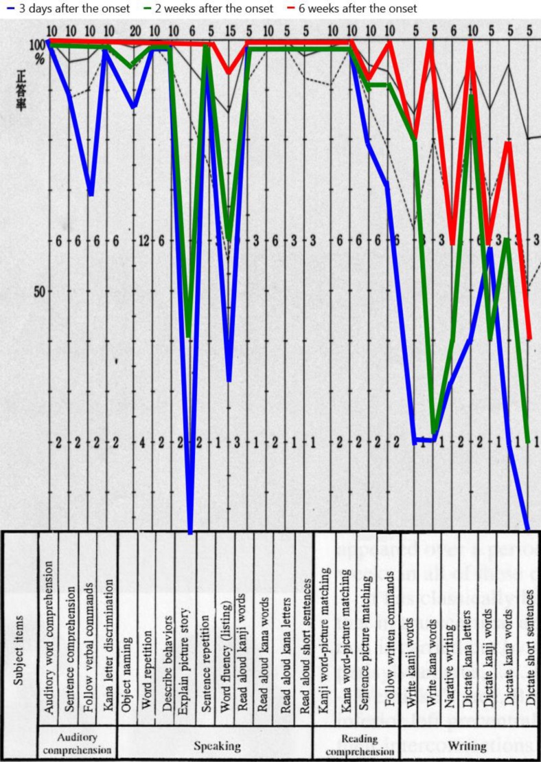
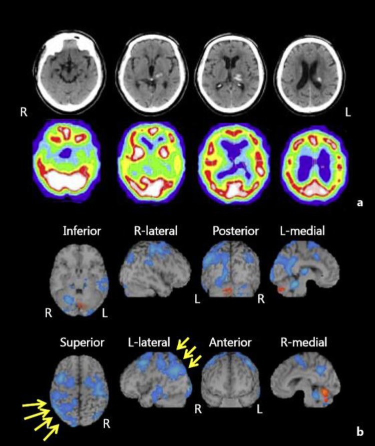
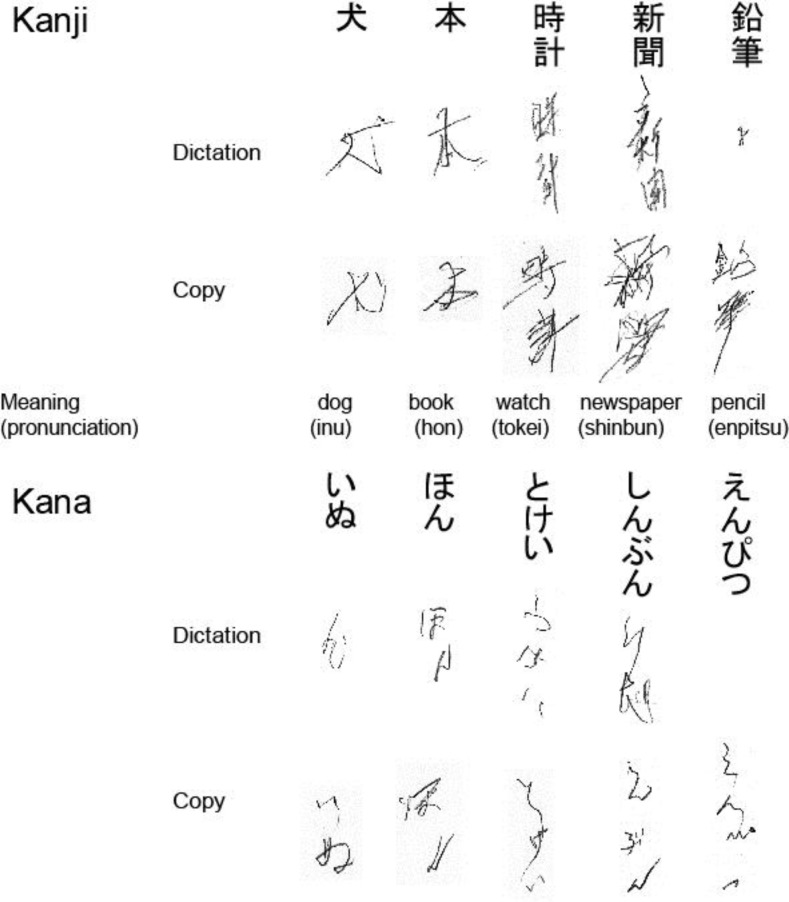
A 71-year-old right-handed man was admitted to our hospital with right hemiparesis and sensory impairment associated with mild aphasia. Although aphasia gradually resolved within 2 weeks after stroke onset, his writing ability remained disturbed. A computed tomography (CT) scan at stroke onset revealed a hematoma in the left thalamus, but no cortical lesions were observed. Further, a single-photon emission CT (SPECT) scan showed decreased blood flow in the left thalamus, in the cortical region extending from the left superior temporal gyrus to the parietal lobe, and in the frontal lobe. It is possible that agraphia may have directly resulted from the thalamic lesion, but SPECT findings strongly suggested that a general decrease in left cortical function concomitant with a disruption of the thalamocortical and cortico-thalamocortical projection fibers produced these cognitive deficits.
Keywords: Agraphia; Aphasia; Cerebral hemorrhage; Stroke.
Figures
Similar articles
-
Thalamic alexia with agraphia.Neurol Int. 2012 Jan 9;4(1):e4. doi: 10.4081/ni.2012.e4. Epub 2012 Feb 9. Neurol Int. 2012. PMID: 22593808 Free PMC article.
-
Functional dissociation between Kana and Kanji: agraphia following a thalamic hemorrhage.Neurol Sci. 2012 Apr;33(2):409-13. doi: 10.1007/s10072-011-0753-7. Epub 2011 Sep 6. Neurol Sci. 2012. PMID: 21894554
-
[A case of progressive multifocal leukoencephalopathy presenting white matter MRI lesions extending over the cerebral cortex and a marked decrease in cerebral blood flow on SPECT, and associated with HTLV-I infection].Rinsho Shinkeigaku. 2005 Jun;45(6):426-30. Rinsho Shinkeigaku. 2005. PMID: 16022467 Review. Japanese.
-
["Left unilateral agraphia with right hemiparesis" after occlusion of the left middle cerebral artery].No To Shinkei. 1992 Jul;44(7):661-6. No To Shinkei. 1992. PMID: 1419344 Japanese.
-
Global aphasia due to thalamic hemorrhage: a case report and review of the literature.Arch Phys Med Rehabil. 1996 Dec;77(12):1312-5. doi: 10.1016/s0003-9993(96)90199-9. Arch Phys Med Rehabil. 1996. PMID: 8976318 Review.
Cited by
-
Cerebral activations related to writing and drawing with each hand.PLoS One. 2015 May 8;10(5):e0126723. doi: 10.1371/journal.pone.0126723. eCollection 2015. PLoS One. 2015. PMID: 25955655 Free PMC article.
-
Voxel-based lesion analysis of brain regions underlying reading and writing.Neuropsychologia. 2018 Jul 1;115:51-59. doi: 10.1016/j.neuropsychologia.2018.03.021. Epub 2018 Mar 20. Neuropsychologia. 2018. PMID: 29572061 Free PMC article.
-
Aphasia and unilateral spatial neglect due to acute thalamic hemorrhage: clinical correlations and outcomes.Neurol Sci. 2016 Apr;37(4):565-72. doi: 10.1007/s10072-016-2476-2. Epub 2016 Jan 21. Neurol Sci. 2016. PMID: 26796359
References
-
- Hillemand P: Contribution à l’étude des syndromes thalamiques; thèse, Paris, 1925.
-
- Lhermitte J. Symptomatologie de l'hémorragie du thalamus. Rev Neurol (Paris) 1936;65:89–93.
-
- Fisher CM. The pathologic and clinical aspects of thalamic hemorrhage. Trans Am Neurol Assoc. 1959;84:56–59. - PubMed
-
- Alexander MP, LoVerme SR., Jr Aphasia after left hemispheric intracerebral hemorrhage. Neurology. 1980;30:1193–1202. - PubMed
-
- Toyokura M, Kobayashi R, Aono KA. A case of pure agraphia due to left thalamic hemorrhage. Tokai J Exp Clin Med. 2010;35:89–94. - PubMed
Publication types
LinkOut - more resources
Full Text Sources
Other Literature Sources
