. 2013 Apr 19;8(4):e61270.
doi: 10.1371/journal.pone.0061270. Print 2013.

The Drosophila BTB domain protein Jim Lovell has roles in multiple larval and adult behaviors

Affiliations

The Drosophila BTB domain protein Jim Lovell has roles in multiple larval and adult behaviors

Sonia M Bjorum et al. PLoS One. .

Erratum in

  • PLoS One. 2013;8(8). doi:10.1371/annotation/88d89372-9fdc-48d7-83b3-26b61142a5e2

Abstract

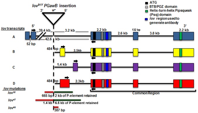
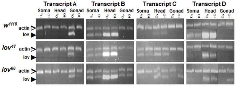
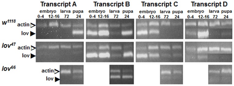
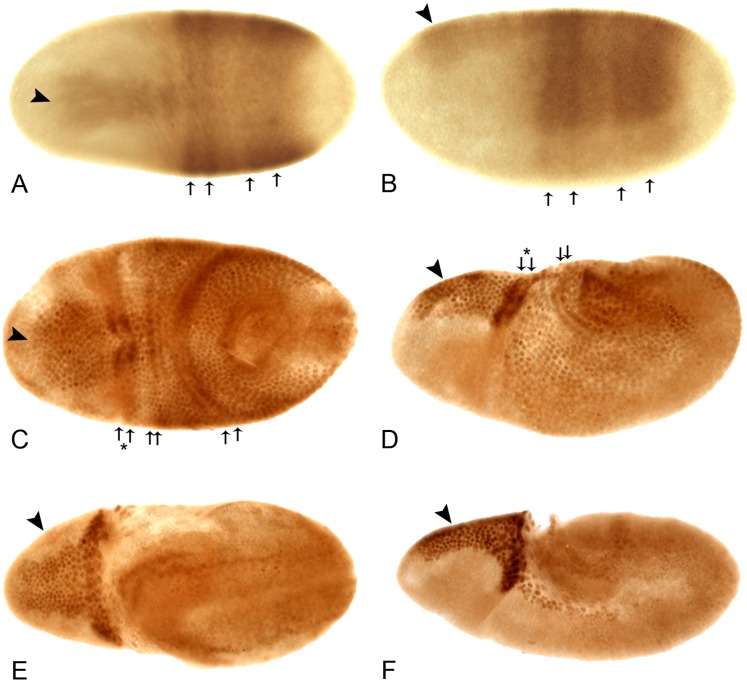
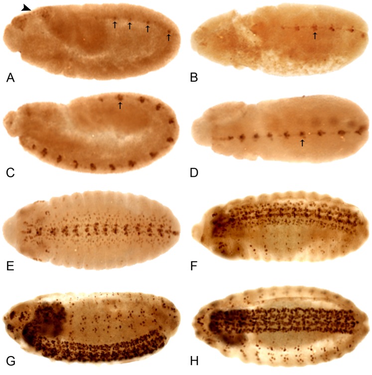
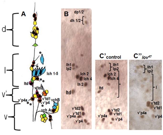
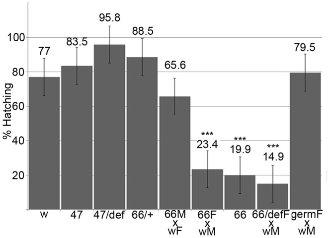
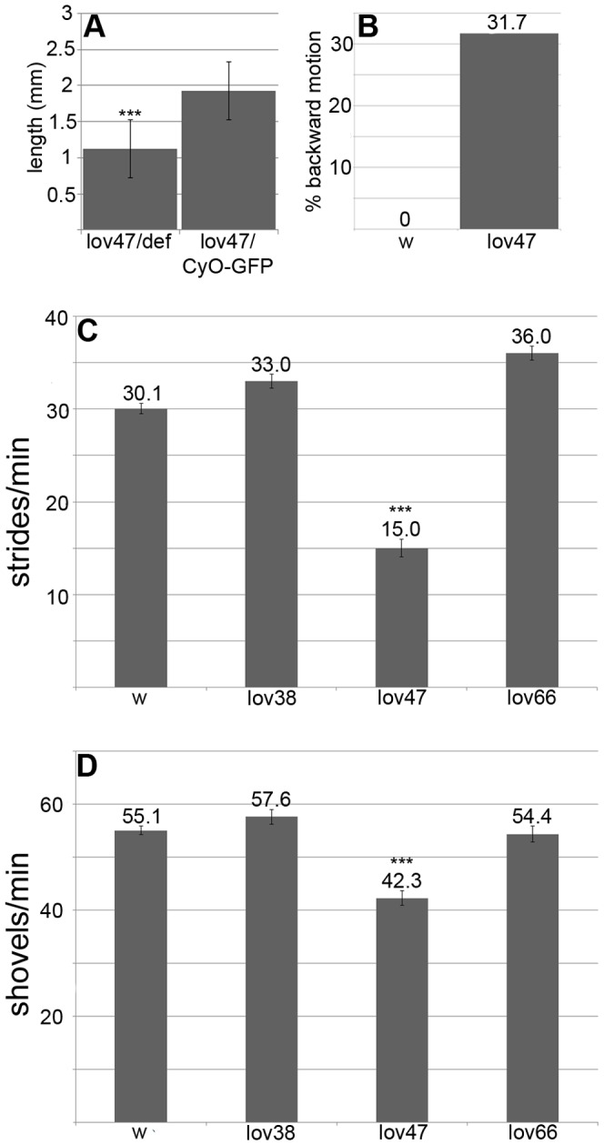
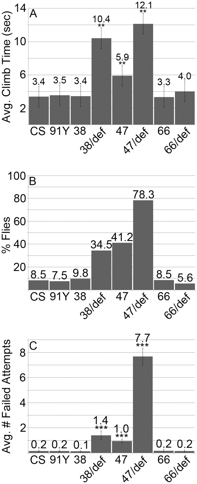
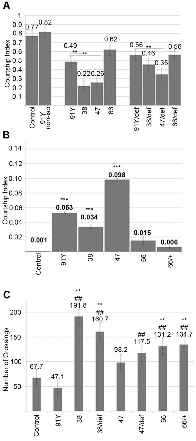
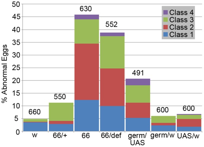
Innate behaviors have their origins in the specification of neural fates during development. Within Drosophila, BTB (Bric-a-brac,Tramtrack, Broad) domain proteins such as Fruitless are known to play key roles in the neural differentiation underlying such responses. We previously identified a gene, which we have termed jim lovell (lov), encoding a BTB protein with a role in gravity responses. To understand more fully the behavioral roles of this gene we have investigated its function through several approaches. Transcript and protein expression patterns have been examined and behavioral phenotypes of new lov mutations have been characterized. Lov is a nuclear protein, suggesting a role as a transcriptional regulator, as for other BTB proteins. In late embryogenesis, Lov is expressed in many CNS and PNS neurons. An examination of the PNS expression indicates that lov functions in the late specification of several classes of sensory neurons. In particular, only two of the five abdominal lateral chordotonal neurons express Lov, predicting functional variation within this highly similar group. Surprisingly, Lov is also expressed very early in embryogenesis in ways that suggests roles in morphogenetic movements, amnioserosa function and head neurogenesis. The phenotypes of two new lov mutations that delete adjacent non-coding DNA regions are strikingly different suggesting removal of different regulatory elements. In lov(47) , Lov expression is lost in many embryonic neurons including the two lateral chordotonal neurons. lov(47) mutant larvae show feeding and locomotor defects including spontaneous backward movement. Adult lov(47) males perform aberrant courtship behavior distinguished by courtship displays that are not directed at the female. lov(47) adults also show more defective negative gravitaxis than the previously isolated lov(91Y) mutant. In contrast, lov(66) produces largely normal behavior but severe female sterility associated with ectopic lov expression in the ovary. We propose a negative regulatory role for the DNA deleted in lov(66) .


Conflict of interest statement

Competing Interests: The authors have declared that no competing interests exist.

Figures

Figure 1
Figure 1. Transcripts and mutations of the Drosophila jim lovell locus.
The exon structure of the four lov transcripts is shown. The three common exons encode the same BTB/POZ domain protein. Structural motifs of the protein and the region used to prepare antibodies are indicated. Arrows indicate the positions of the primers used to probe for expression of the four transcripts. The original lov91Y P{GawB} insertion and three deletion mutations (lov38, lov47 and lov66) generated by imprecise excision of the transposon are shown.
Figure 2
Figure 2. Tissue specific expression of the lov transcripts in adults.
Semi-Q RT-PCR was used to detect lov transcripts in the bodies (soma), heads and gonads of male and female adults from a control (w1118) strain and lov47 and lov66 mutant lines. Primers as indicated in Figure 1. In all cases, the fragments amplified span intron-exon boundaries to limit detection to mature transcripts. A fragment from the ubiquitous Actin mRNA (Actin at 57B) was amplified in parallel as the control. At least two separate RNA preparations for each tissue were used for transcript quantitation. The PCR fragment for transcript D was very close in size to the Actin PCR fragment. To quantitate this transcript, aliquots of separate transcript D and Actin PCR reactions for each RNA sample were run in parallel in separate gel lanes.
Figure 3
Figure 3. Developmental expression of the lov transcripts.
Semi-Q RT-PCR, as described for Figure 2, was used to detect lov transcripts in embryos 0–4 and 12–16 hours after egg laying (AEL), larvae 72 hours AEL, and pupae 24 hours after pupation from control (w1118) and lov47 and lov66 mutant flies. lov66 is maintained as a balanced (lov66/CyO) stock (see text) and transcripts for homozygous lov66 embryos could not be reliably evaluated. At least two separate RNA preparations for each stage were used for transcript quantitation.
Figure 4
Figure 4. The early embryonic expression pattern of Lov protein.
All embryos are anterior to left. A, C, and E - views of the dorsal surface; B, D, and F - lateral views, dorsal uppermost. At cellular blastoderm (A, B) Lov staining appears across the dorsal midline as an anterior “wedge” (arrowhead) and a “saddle” of two pairs of diffuse stripes (arrows) at ∼10–50% egg length. In early germ band extension (C, D), staining in the wedge intensifies (arrowhead) and the diffuse saddle stripes (arrows) sharpen into paired rows of nuclei spanning the positions of ectodermal folds (see text). A further pair of rows of nuclei staining for Lov across the dorsal midline develops at the edges of the cephalic furrow (arrows and asterisk). The staining in these rows is particularly intense where they traverse the wedge of Lov stain. In late germ band extension (E, F), the wedge staining (arrowheads) can be seen to lie within the procephalic neuroectoderm. Intense staining is also seen in the polyploid nuclei of the amnioserosa at its junction with the cephalic furrow and in its lateral longitudinal extensions. In E, paired rows of Lov staining nuclei are also seen at the periphery of the extending germ band.
Figure 5
Figure 5. The late embryonic expression pattern of Lov protein.
All embryos are anterior to the left. A, C, and G - lateral views, dorsal uppermost; B, D, EH - dorsal views. At the end of germ band extension (A) the early Lov pattern (Figure 4) disappears and a single nucleus at the midline of each parasegment begins to express Lov. As germ band retraction proceeds (BD), more nuclei at the developing CNS midline express Lov. Nuclei in the lateral regions of the CNS (E) the brain lobes (E, F) and along the longitudinal connectives (F) develop staining as germ band retraction is completed and dorsal closure begins. PNS staining is first detected in early dorsal closure (F). At ∼ stage 15, CNS and PNS staining are maximal (G, H) and CNS staining has a mesh-like appearance (H).
Figure 6
Figure 6. Lov expression in the embryonic abdominal PNS.
A - a cartoon of the four clusters (dorsal (d), lateral (l), ventral’ (v′) and ventral (v)) of sensory neurons within each hemisegment of the abdominal segments (redrawn from [2]). Neurons are color coded according to class as follows: orange - external sense organ, pink - tracheal dendrite, blue - chordotonal, yellow - dendritic arborization, green - bipolar dendritic. Key neurons discussed in the text are labeled. Neuronal nuclei expressing Lov (shown by black dots or ovals) were identified in embryos co-stained with anti-Lov and 22C10. Grey dots = putative non-neuronal support cell nuclei that express low levels of Lov. Nomenclature for all neurons as in except that the neurons of the lateral chordotonal organ are labeled lch1-5. B - Actual example of wild type Lov-staining nuclei at stage 15 in clusters d, l, and v′ of a single abdominal hemisegment. C - anti-Lov (brown) and 22C10 (blue) costaining of the lateral chordotonal organ in a control embryo to demonstrate Lov limitation to lch2 and lch4. D - 22C10 staining of a lov47 homozygous embryo demonstrating the presence of all five chordotonal neurons in the lateral chordotonal organ. E′, E′′ - loss of Lov staining in chordotonal organ neurons lch2 and 4 of a lov47 mutant embryo. E′ shows control staining in clusters l and v′. E′′ shows staining in equivalent regions of a lov47 embryo. Loss of staining in td neuron ltd is also seen in this lov47 embryo.
Figure 7
Figure 7. Hatch rates for eggs from lov47, lov66 and related genotypes.
Batches of eggs from stocks or crosses of the genotypes indicated were collected and scored for hatching as described in Material and Methods. w = w1118, 47 = lov47, 66 = lov66, def = lov deficiency chromosome SB1,+ = CyO balancer, germ = UAS-lov in the ovarian germline under the GAL4 driver P (GAL4::VP16-nos.UTR) CG6325MVD1. M = male, F = female. Hatch rates for the lov66/CyO stock and the lov47/def stock were adjusted to correct for death of CyO/CyO and def/def homozygotes. At least six separate collections, totaling at least 600 eggs, were scored for each genotype. One way Anova and a Dunnett’s test were used to determine statistical significance *** = p<0.001 compared to w1118.
Figure 8
Figure 8. Growth and behavioral defects in lov47 larvae.
A - poor growth of lov47 larvae in rich medium. lov47 hemizygous (lov47/def) and heterozygous (lov47/CyO-GFP) larvae from a lov47/CyO-GFP stock grown on yeast paste were sorted based on GFP fluorescence at 87 hours AEL and imaged under a dissecting microscope with a length gauge, after brief ether anesthetization. B - percentage of larvae at 72 hours AEL showing backward locomotion (as defined in Material and Methods) for lov47 and w1118 (w). C - forward locomotion rates at 72 hours AEL for w1118, lov38, lov47, and lov66 homozygous larvae. D - food shoveling rates at 72 hours AEL for larval genotypes as in C. Statistics as for Figure 7. *** = significantly decreased (p<0.001) as compared to w1118.
Figure 9
Figure 9. Negative gravitactic defects in lov38 and lov47 adults.
A - Climb times in the negative gravitactic climb assay (see Material and Methods) for flies of each genotype that completed the climb in less than one minute. B - Percentage of flies of each genotype that failed to complete the climb in at least one trial. C - The average number of failed climbs for the 10 consecutive trials performed for each fly. CS = Canton-S, 91Y = lov91Y, 38 = lov38, 47 = lov47, 66 = lov66, def - lov deficiency chromosome SB1. Statistics as previously. ** = p<0.01, *** = p<0.001 as compared to CS.
Figure 10
Figure 10. Courtship defects in lov38 and lov47 males.
A - Directed courtship. Courtship indices for courtship directed towards the female. The courtship indices for lov91Y in its original genetic background (91Y non-iso) and after isogenization of lov91Y into the same w+ background (see text) as the other lov mutants (91Y) are shown. Comparison of these two indices demonstrates that the new genetic background suppresses courtship significantly. Courtship behavior for lov38, lov47 and lov66 is therefore compared only to the lov91Y iso line (91Y) to correct for this background suppression of courtship. Statistics as previously. ** = p<0.01 as compared to isogenized lov91Y. B - Non-directed courtship. Courtship indices for elements of the courtship ritual performed while not pursuing the female. Statistics as previously. *** = p<0.001 as compared to Canton-S control. C - Locomotor activity for males alone in courtship chambers (see Material and Methods). Control - Canton-S, 91Y = lov91Y, 38 = lov38, 47 = lov47, 66 = lov66,+ = CyO chromosome, def - lov deficiency chromosome SB1. Statistics as previously. ** = p<0.01 as compared to Canton-S control. ## = p<0.01 as compared to isogenized lov91Y.
Figure 11
Figure 11. Classes of abnormal eggs produced by lov66 mothers.
A - wild type egg with opaque chorion and two opaque dorsal appendages (DAs). B-F - examples of abnormal eggs from lov66 homozyous or hemizygous mothers. B - class 1 egg (see text) with translucent chorion and translucent, weak DAs. C - class 2 egg (see text). DAs are abnormal and translucent and formation of the anterior chorion has failed. Illuminated to show chorion-like barrier (arrow) that has prevented complete nurse cell dumping. D - class 3 egg. Chorion is opaque and thick but egg is short, with stubby DAs. E, F - class 2 short, flaccid eggs with translucent chorions and translucent abnormal DAs.
Figure 12
Figure 12. Abnormal egg production in lov66-related genotypes.
The percentage of abnormal eggs of various classes (see text) is shown for lov66 homozygous (66), heterozygous (66/+ = 66/CyO), and hemizygous mothers (66/def), for mothers (germ/UAS) expressing UAS-lov ectopically in the ovarian germ-line under the GAL4 driver P (GAL4::VP16-nos.UTR) CG6325MVD1 and for appropriate control mothers (w = w1118, germ/w and UAS/w). All females were mated to w1118 males. Numbers of eggs examined for each genotype are shown above the bars.

Similar articles

Cited by

References

    1. Bertrand N, Castro DS, Guillemot F (2002) Proneural genes and the specification of neural cell types. Nature Reviews in Neuroscience 3: 517–530. - PubMed
    1. Ghysen A, Dambly-Chaudière C (1993) The specification of sensory neuron identity in Drosophila. BioEssays 15: 293–298. - PubMed
    1. Kearney JB, Wheeler SR, Estes P, Parente B, Crews ST (2004) Gene expression profiling of the developing Drosophila CNS midline cells. Developmental Biology 275: 473–492. - PMC - PubMed
    1. Wheeler SR, Kearney JB, Guardiola AR, Crews ST (2006) Single-cell mapping of neural and glial gene expression in the developing Drosophila CNS midline cells. Developmental Biology 294: 509–524. - PMC - PubMed
    1. Wheeler SR, Stagg SB, Crews ST (2008) Multiple Notch signaling events control Drosophila CNS midline neurogenesis, gliogenesis and neuronal identity. Development 135: 3071–3079. - PMC - PubMed

Publication types

MeSH terms