. 2013 Apr 19;8(4):e61340.
doi: 10.1371/journal.pone.0061340. Print 2013.

Spatio-temporal distribution of Smads and role of Smads/TGF-β/BMP-4 in the regulation of mouse bladder organogenesis

Affiliations

Spatio-temporal distribution of Smads and role of Smads/TGF-β/BMP-4 in the regulation of mouse bladder organogenesis

Syed S Islam et al. PLoS One. .

Abstract

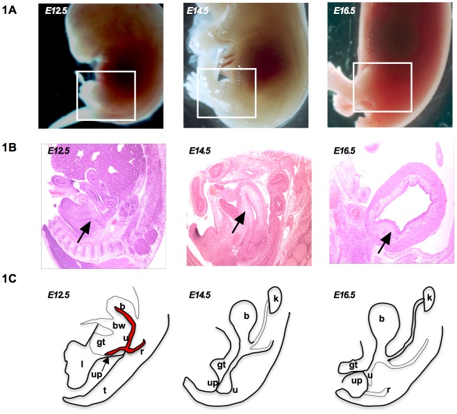
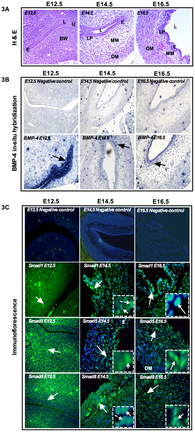
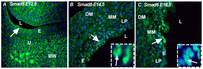
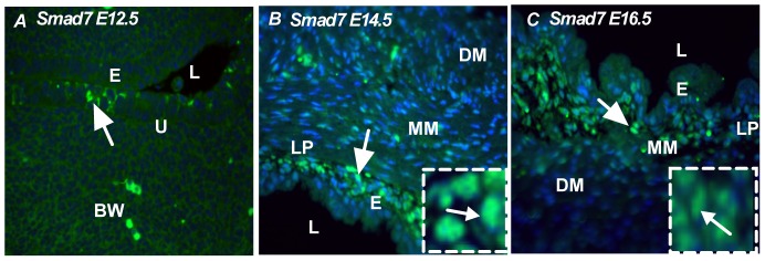
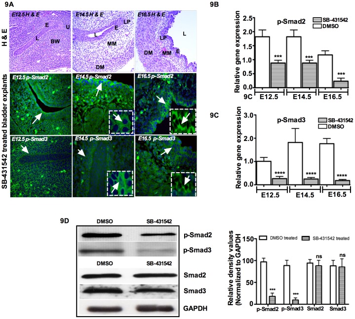
Although Shh, TGF-β and BMP-4 regulate radial patterning of the bladder mesenchyme and smooth muscle differentiation, it is not known what transcription factors, local environmental cues or signaling cascades mediate bladder smooth muscle differentiation. We investigated the expression patterns of signaling mediated by Smad2 and Smad3 in the mouse embryonic bladder from E12.5 to E16.5 by using qRT-PCR, in situ hybridization and antibodies specifically recognizing individual Smad proteins. The role of Smad2 and Smad3 during smooth muscle formation was examined by disrupting the Smad2/3 signaling pathway using TβR1 inhibitor SB-431542 in organ culture system. qRT-PCR results showed that R-Smads, Co-Smad and I-Smads were all expressed during bladder development. RNA ISH for BMP-4 and immunostaining of TGF-β1 showed that BMP-4 and TGF-β1 were expressed in the transitional epithelium, lamina propia and muscularis mucosa. Smad1, Smad5 and Smad8 were first expressed in the bladder epithelium and continued to be expressed in the transitional epithelium, muscularis mesenchyme and lamina propia as the bladder developed. Smad2, Smad3 and Smad4 were first detected in the bladder epithelium and subsequently were expressed in the muscularis mesenchyme and lamina propia. Smad6 and Smad7 showed overlapping expression with R-Smads, which are critical for bladder development. In bladder explants (E12.5 to E16.5) culture, Smad2 and Smad3 were found localized within the nuclei, suggesting critical transcriptional regulatory effects during bladder development. E12.5 to E16.5 bladders were cultured with and without TβR1 inhibitor SB-431542 and assessed by qRT-PCR and immunofluorescence. After three days in culture in SB-431542, α-SMA, Smad2 and Smad3 expressions were significantly decreased compared with controls, however, with no significant changes in the expression of smooth muscle myosin heavy chain (SM-Myh. Based on the Smad expression patterns, we suggest that individual or combinations of Smads may be necessary during mouse bladder organogenesis and may be critical mediators for bladder smooth muscle differentiation.


Conflict of interest statement

Competing Interests: The authors have declared that no competing interests exist.

Figures

Figure 1
Figure 1. Schematic of depicting urogenital organs adjacent to the limb and the tail at E12.5 to E16.5.
A) Whole embryos from E12.5 to E16.5 B) H & E sections from E12.5 to E16.5 and C) Bladder developmental progression. b: bladder, bw: body wall, u: urethra, gt: genital tubercle, up: urethral plate, r: rectuml, limb, t: tail, k: kidney.
Figure 2
Figure 2. qRT-PCR analysis of the relative expression levels of TGF-β1, BMP-4 and Smads in embryonic bladders from E12.5 to neonatal day0.
Ten embryonic bladders per group were lysed to extract total RNA for qRT-PCR. Data were normalized to that of GAPDH and the relative expression levels are shown. Each experiment was repeated three times and the mean value was calculated as expression value for each stage of embryonic bladder development. A) BMP-responsive R-Smads. Smad1, Smad5 and Smad8 were expressed in all gestation stages. Smad1 expression was minimal in E12.5 and Smad8 expression was low in E18.5 and neonatal day0 (Mann-Whitney test, p<0.005) B) TGF-β responsive R-Smads. Smad2 and Smad3 showed relatively high expression in all gestation stages while Smad2 showed decreased expression at E18.5 and neonatal day0. (Mann-Whitney test, p<0.005) C) Common Smad. Smad4 showed the highest expression pattern from E12.5 to E16.5 and decreased in E18.5 and neonatal day0 (Mann-Whitney test, p<0.005) and D) Inhibitory Smads. Smad6 and Smad7. Smad6 showed higher expression from E12.5 to E16.5, and lowest expression levels in E18.5 and neonatal day0 (Mann-Whitney test, p<0.005), while Smad7 showed the highest expression level at E12.5 and decreased in the subsequent stages (Mann-Whitney test, p<0.005).
Figure 3
Figure 3. Expression pattern of BMP-4 and BMP-responsive Smads, Smad1, Smad5 and Smad8.
A) Top panel shows H & E staining of mouse bladder anatomy from E12.5 to E16.5. B) In situ hybridization of BMP-4 (middle panel). Negative controls of BMP-4 (top middle panel). No BMP-4 expression was detected for negative control from E12.5 to E16.5. At E12.5, BMP-4 was localized in the bladder epithelium and urethra (arrow). At E14.5, BMP-4 was localized adjacent to the submucosa (lamina propia) and muscularis mesenchyme and periphery of the detrusor muscle (arrow). At E16.5 BMP-4 expression was similar to E14.5, but expression level declined (arrow). C) Immunofluorescence staining of Smad1, Smad5 and Smad8 (bottom panel). Negative control staining sections were incubated without primary antibodies (top bottom panel). At E12.5, Smad1, Smad5 and Smad8 were all localized in the bladder epithelium and body wall and urethra. At E14.5 Smad1 was localized in the transitional epithelium and muscularis mesenchyme and less intensely in the detrusor muscle (arrow). Insert: positive staining of phosphorylated Smad1. At E14.5, Smad5 and Smad8 showed similar expression pattern to Smad1. Both Smads were localized in the transitional epithelium and expressed in the muscularis mesenchyme (arrow). Insert: positive staining of phosphorylated Smad5 and Smad8. At E16.5, Smad1 and Smad5 showed strong expression in the transitional epithelium and lamina propia and sporadic expression in muscularis mesenchyme, but Smad8 ocalized less intensely in the transitional epithelium and muscularis mucosa (arrow). Magnification ×40. e: epithelum, l: lumen, bw: body wall, u: urethra, lp: lamina propia, mm: muscularis mesenchyme, dm: detrusor muscle. (Blue color: DAPI, Green color: Respective Smads).
Figure 4
Figure 4. Expression pattern of TGF-β1 and TGF-β responsive Smads, Smad2 and Smad3.
A) Top left panel shows H & E staining of mouse bladder anatomy from E12.5 to E16.5. B) Negative control sections of TGF-β1 and C) Smad2 and Smad3 incubated only with secondary antibodies (no primary antibodies, top middle and right panel). D) At E12.5, TGF-β1 expression was faint and localized in the epithelium and urethra (bottom left panel). At E14.5 and E16.5, TGF-β1 was localized in the transitional epithelium, adjacent to the lamina propia and muscularis mesenchyme (arrow). E and F) At E12.5, Smad2 and Smad3 were in the bladder epithelium and urethra (arrow, bottom middle and right panel). At E14.5, Smad2 and Smad3 were localized in the transitional epithelium, lamina propia and muscularis mucosa (arrow). Insert: positive staining of phosphorylated Smad2 and Smad3 (bottom middle right panel). At E16.5, Smad2 was localized in the transitional epithelium and periphery of the detrusor muscle (arrow, bottom middle panel) and Smad3 was localized in the transitional epithelium, muscularis mesenchyme and periphery of the detrusor muscle (arrow, bottom right panel). Insert: positive staining of phosphorylated Smad2 and Smad3. Magnification ×40. e: epithelum, l: lumen, bw: body wall, u: urethra, lp:- lamina propia, mm: muscularis mesenchyme, dm: detrusor muscle. (Green color: Respective Smads, Blue color: DAPI).
Figure 5
Figure 5. Expression pattern of common Smad, Smad4.
A) At E12.5, Smad4 was localized in the bladder epithelium and urethra (arrow). B) At E14.5, Smad4 was localized in the transitional epithelium, muscularis mesenchyme, lamina propia and detrusor muscle (arrow) Insert: nuclear localization of Smad4. C) At E16.5, Smad4 was localized in the inner epithelium, less intensely in the muscularis mesenchyme and detrusor muscle (arrow) Insert: nuclear localization of Smad4. Magnification ×40. e: epithelum, l: lumen, bw: body wall, u: urethra, lp: lamina propia, mm: muscularis mesenchyme, dm: detrusor muscle. (Green color: Smad4, Blue color: DAPI).
Figure 6
Figure 6. Expression pattern of inhibitory Smad, Smad6.
A) At E12.5, Smad6 was localized in the bladder epithelium (arrow). B) At E14.5, Smad6 was localized in the transitional epithelium (arrow). C) At E16.5, Smad6 was predominantly localized in the transitional epithelium and less intensely in the muscularis mesenchyme (arrow) Insert: nuclear localization of Smad6. Magnification ×40. e: epithelum, l: lumen, bw: body wall, u: urethra, lp: lamina propia, mm: muscularis mesenchyme, dm: detrusor muscle. (Green color: Smad6, Blue color: DAPI).
Figure 7
Figure 7. Expression pattern of inhibitory Smad, Smad7.
A) At E12.5, Smad7 was less intense and localized in the bladder epithelium (arrow). B) At E14.5, Smad7 was localized in the transitional epithelium, lamina propia (arrow) Insert: Nuclear localization of Smad7. C) At E16.5, Smad7 was predominantly localized in lamina propia and less intensely in the muscularis mucosa (arrow) Insert: nuclear localization of Smad7. Magnification ×40. e: epithelum, l: lumen, bw: body wall, u: urethra, lp: lamina propia, mm: Muscularis mesenchyme, dm: detrusor muscle. (Green color: Smad7, Blue color: DAPI).
Figure 8
Figure 8. Effects of SB-431542 in bladder smooth muscle cell differentiation: A) Top left panel.
E12.5, E14.5 and E16.5 bladders were cultured in DMEM/F12 50∶50 supplemented with insulin-transferrin and penicillin/streptomycin antibiotics. Bladder explants cultures treated for three days in DMSO only and showing H & E staining sections. (Phase contrast microscope Magnification 4×). D) Top right panel: Bladder explants culture treated in TβRI inhibitor SB-431542 for three days and showing with H & E staining section. (Phase contrast microscope Magnification 4×). B and C) α-SMA and SM-Myh mRNA gene expression against GAPDH by qRT-PCR at E12.5 to E16.5 in cultured embryonic bladders after three days of organ culture (middle panel). E) Immunofluorescence staining of α-SMA and SM-Myh and expressed at embryonic days from E14.5 to E16.5 in the untreated bladder explants (middle top panel) and α-SMA expression was faint at E14.5 to E16.5 after three days treatment with SB-431542 (top right panel). F and H) In contrast, SM-Myh expression showed no changes in the untreated and treated group (bottom left and right panel). I) Western blot analysis showing the expression of α-SMA and SM-Myh with densitometry analysis. In SB-431542 treated bladder explants, α-SMA was decreased by 90%, but no significant changes were observed in SM-Myh. e: epithelium, l: lumen, bw: body wall, u: urethra, lp: lamina propia, mm: muscularis mesenchyme, dm: detrusor muscle. (Green color: α-SMA and SM-Myh, Blue color: DAPI).
Figure 9
Figure 9. Induction of α-SMA by TGF-β in cultured bladder organ requires Smad2 and Smad3.
E12.5, E14.5 and E16.5 bladders were cultured in DMEM/F12 50∶50 supplemented with insulin-transferrin and penicillin/streptomycin antibiotics. A) Top panel: H & E staining of bladder anatomy from E12.5 to E16.5. Blockade of Smad2 and Smad3 by SB-431542 in cultured bladder as detected by immunofluorescence. Cultured bladders were treated with SB-431542 for three days and processed for immunofluorescence using p-Smad2 and p-Smad3 antibodies. Insert: staining of phosphorylated Smad2 and Smad3. Nuclei of the same cells stained with DAPI. B and C) p-Smad2 and p-Smad3 mRNA gene expression against GAPDH by qRT-PCR at E12.5 to E16.5 in cultured embryonic bladders after three days with and without SB-431542. D) Western blot analysis showing the expression of p-Smad2 and p-Smad3 with densitometry analysis. In SB-431542 treated bladder explants, p-Smad2 was decreased by 85% and p-Smad3 was decreased by more than 95%. No changes were observed for total Smad2 and Smad3 expression. Magnification ×40. e: epithelum, l: lumen, bw: body wall, u: urethra, lp: lamina propia, mm: muscularis mesenchyme, dm: Detrusor muscle. (Green color: Respective Smads, Blue color: DAPI).
Figure 10
Figure 10. Expression pattern summary of Smads.
ISH and Immunofluorescence staining revealed expression pattern of BMP-4, TGF-β1 and Smads based on intensity of expression for particular Smads within the bladder cellular compartment at E14.5 and six different kinds of expression pattern summarized below: A) Sagittal section of mouse bladder. B) BMP-4 was strongly expressed in the muscularis mesenchyme, medium in the lamina propia and weaker in the epithelium and detrusor muscle. C) Smad1, Smad5 and Smad8 were highly expressed in the bladder epithelium and muscularis mesenchyme, medium level in the lamina propia and weaker expression in the detrusor muscle. D) TGF-β1 highly expressed in the lamina propia and muscularis mesenchyme and weaker expression in epithelium and detrusor muscle. E) Smad2 and Smad3 were highly expressed in the epithelium and muscularis mesenchyme, medium level in the lamina propia and weaker expression in the detrusor muscle. F) Smad4 expression level was high in the epithelium, lamina propia, muscularis mesenchyme and detrusor muscle respectively and G) Smad6 and Smad7 were highly expressed in the epithelium but medium level in the lamina propia and weaker expression in the muscularis mesenchyme and detrusor muscle.

Similar articles

Cited by

References

    1. Li J, Shiroyanagi Y, Lin G, Haqq C, Lin CS, et al. (2006) Serum response factor, its cofactors, and epithelial-mesenchymal signaling in urinary bladder smooth muscle formation. Differentiation 74: 30–39. - PubMed
    1. Shiroyanagi Y, Liu B, Cao M, Agras K, Li J, et al. (2007) Urothelial sonic hedgehog signaling plays an important role in bladder smooth muscle formation. Differentiation 75: 968–977. - PubMed
    1. Baskin LS, Hayward SW, Young P, Cunha GR (1996) Role of mesenchymal-epithelial interaction in normal bladder development. J Urol 156: 1820–1827. - PubMed
    1. Baskin LS, Sutherland RA, DiSandro MS, Thompson AA, Cunha GR (1997) Cellular signaling in the bladder. Frontiers in Bioscience 2: 592–595. - PubMed
    1. Baskin LS, Hayward SW, Young P, Cunha GR (1996a) Role of mesenchymal-epithelial interaction in bladder development. J Urol 156: 1820–1827. - PubMed

MeSH terms