. 2013 Apr 19;8(4):e61400.
doi: 10.1371/journal.pone.0061400. Print 2013.

Chlamydia trachomatis infection induces replication of latent HHV-6

Affiliations

Chlamydia trachomatis infection induces replication of latent HHV-6

Bhupesh K Prusty et al. PLoS One. .

Abstract

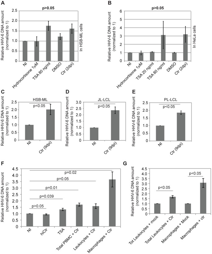
Human herpesvirus-6 (HHV-6) exists in latent form either as a nuclear episome or integrated into human chromosomes in more than 90% of healthy individuals without causing clinical symptoms. Immunosuppression and stress conditions can reactivate HHV-6 replication, associated with clinical complications and even death. We have previously shown that co-infection of Chlamydia trachomatis and HHV-6 promotes chlamydial persistence and increases viral uptake in an in vitro cell culture model. Here we investigated C. trachomatis-induced HHV-6 activation in cell lines and fresh blood samples from patients having Chromosomally integrated HHV-6 (CiHHV-6). We observed activation of latent HHV-6 DNA replication in CiHHV-6 cell lines and fresh blood cells without formation of viral particles. Interestingly, we detected HHV-6 DNA in blood as well as cervical swabs from C. trachomatis-infected women. Low virus titers correlated with high C. trachomatis load and vice versa, demonstrating a potentially significant interaction of these pathogens in blood cells and in the cervix of infected patients. Our data suggest a thus far underestimated interference of HHV-6 and C. trachomatis with a likely impact on the disease outcome as consequence of co-infection.


Conflict of interest statement

Competing Interests: The authors have declared that no competing interests exist.

Figures

Figure 1
Figure 1. Chlamydia trachomatis infection induces CiHHV-6 DNA replication.
(A) qPCR analysis of viral DNA in HSB-ML cells after 2 days of C. trachomatis infection. HSB-ML cells were infected with C. trachomatis (Ctr) for 2 days. In parallel, the same number of HSB-ML cells was cultured in the presence of 1 µM hydrocortisone or 80 ng/ml of TSA for 2 days. As TSA was dissolved in DMSO, a DMSO control was also included in the study. Data represent the mean ± SEM of 5 independent experiments. (B) qPCR analysis of viral DNA in HeLa cells carrying latent HHV-6A after 2 days of C. trachomatis infection. Cells were infected with C. trachomatis or were treated with TSA or hydrocortisone as mentioned in (A). Data represent the mean ± SEM of 5 independent experiments. (C, D, E) qPCR analysis of viral DNA in HSB-ML (C), JL-LCL (D) and PL-LCL (E) cells after 9 days of C. trachomatis infection. Cells were infected with C. trachomatis at an MOI of 2–5 for 2 days after which cells were washed thoroughly and grown in presence of fresh media with 1 µg/ml of doxycycline for another 7 days. Total DNA was isolated and used for qPCR assay. Data represent the mean ± SEM of three independent experiments. (F) qPCR analysis of viral DNA in freshly isolated blood cells from CiHHV-6 individuals after 5 days of C. trachomatis infection. Fresh PBMCs were isolated from total blood and were infected with C. trachomatis for 2 days at an MOI of 5 after which cells were washed thoroughly and grown in presence of fresh media with 1 µg/ml of doxycycline for another 3 days. In parallel, the same numbers of PBMCs were cultured in presence of 80 ng/ml of TSA for 5 days. Total leukocytes were separated from monocyte-derived macrophages and were infected with C. trachomatis in parallel. Heat inactivated C. trachomatis (hiCtr) were added to total PBMCs in a separate well and were included as negative control. Data represent the mean ± SEM of results from blood samples of 5 different CiHHV-6 individuals. (G) qPCR analysis of viral DNA in total leukocytes and monocyte-derived macrophages from CiHHV-6 individuals after 5 days of C. trachomatis infection. Total leukocytes were separated from monocyte-derived macrophages and were infected with C. trachomatis for 2 days at an MOI of 5 after which cells were washed thoroughly and grown in presence of fresh media with 1 µg/ml of doxycycline for another 3 days. Data represent the mean ± SEM of three independent experiments performed at the same time from one CiHHV-6 individual.
Figure 2
Figure 2. C. trachomatis infection induces formation of extra-chromosomal HHV-6 DNA in CiHHV-6 cell lines and patient blood samples.
(A) C. trachomatis infection induces viral DNA replication in HSB-ML cells. HSB-ML cells were infected with C. trachomatis at an MOI of 2 for 2 days after which cells were washed thoroughly and grown in presence of fresh media with 1 µg/ml of doxycycline for 35 days. Total DNA was isolated at 3 different time intervals and used for qPCR assay. Data represent the mean ± SEM of 3 independent experiments. (B) Southern hybridization of C. trachomatis-infected HSB-ML cells shows detectable amount of extra-chromosomal HHV-6 after 35 days of infection. Total DNA from non-infected (NI) and 35 days post infected (dpi) HSB-ML cells were run on 1% agarose gel and were subsequently used for southern transfer and hybridization. HHV-6 specific band is marked by dark arrowhead. (C) Ethidium bromide (EtBr) stained agarose gel showing full-length HHV-6 DNA in total PBMCs of a CiHHV-6 individual after 16 days of Chlamydia treatment. Fl, full-length. (D) Same gel as in (C) was processed for neutral-neutral 2D DNA electrophoresis and subsequent hybridization with a HHV6 probe and then with C. trachomatis probe. Migration rate of full-length HHV-6 DNA from C. trachomatis-infected cells is different from that of mock infected cells. Ori, the position of the origin of the second-dimension gel electrophoresis. The positions of the size markers for the first-dimension gel electrophoresis are shown at the top. (E) Gardella gel analysis and subsequent southern hybridization showing extra-chromosomal HHV-6 DNA after C. trachomatis infection. HSB-ML and JL-LCL cells were infected with C. trachomatis (MOI 5) for 2 days after which cells were washed thoroughly and grown in presence of fresh media with 1 µg/ml of doxycycline for another 7 days. Fresh HSB-2 cells were added to these cells and were co-cultured for another 2 weeks. 2.5×10⁶ of these cells were loaded directly onto a Gardella gel and subsequently stained with ethidium bromide and afterwards transferred to a nylon membrane. HSB-2, HHV-6-infected HSB-2 for productive virus infection (HSB-2+HHV-6), C. trachomatis elementary bodies (Ctr EB) and C. trachomatis-infected HSB-ML cells, which were subsequently not co-cultured with HSB-2 cells (HSB-ML+Ctr) were loaded as controls. Nylon membrane was hybridized first with a HHV-6-specific probe and subsequently stripped and used again for hybridization with C. trachomatis-specific probe. Extra-chromosomal linear HHV-6 DNA is marked with *and possible nicked circular viral DNA is marked with a rectangle. Chlamydial DNA did not run in the gel due to the size of genomic DNA and hence C. trachomatis-specific hybridization was observed only in the wells of lane 4 and 5. A clear band (probably the chlamydial plasmid) in lane 3, which was observed after ethidium bromide staining was not detected by C. trachomatis-specific probe. Instead some additional bands were detected in lane 4 with C. trachomatis-specific probes. (F) Transmission electron microscopy pictures showing appearance of particle like condensed DNA structures (marked by black arrowhead) in CiHHV-6 cell nuclei after Chlamydia infection.
Figure 3
Figure 3. Quantitative real time PCR assay to detect HHV-6 and chlamydial load in cervical swabs and total blood samples.
(A–C) Standard curves for HHV-6 and C. trachomatis quantification. Ten fold serial dilutions of U94 HHV-6A (A), C. trachomatis (B) and PI15 (C) plasmids were tested in triplicates in qPCR reaction. Mean Ct values were plotted against the copy numbers. Correlation coefficients and slope values are mentioned in each figure. (D) Scatter plot showing quantitative distribution of HHV-6 and C. trachomatis in cervical swabs of patients (n = 65) with suspected C. trachomatis infection as determined by real-time qPCR assay. Samples with no detectable HHV-6 and C. trachomatis are not included. Samples with HHV-6 DNA below detection limit (5 copies/103 cells) were arbitrarily specified as 4 copies/103 cells. Similarly, samples with less than 100 copies/103 cells were arbitrarily specified as 50 copies/103 cells. Sensitivity of PCR was marked as detection limits.

Similar articles

Cited by

References

    1. Lindquester GJ, Pellett PE (1991) Properties of the human herpesvirus 6 strain Z29 genome: G+C content, length, and presence of variable-length directly repeated terminal sequence elements. Virol 182: 102–110. - PubMed
    1. Chen M, Popescu N, Woodworth C, Berneman Z, Corbellino M, et al. (1994) Human herpesvirus 6 infects cervical epithelial cells and transactivates human papillomavirus gene expression. J Virol 68: 1173–1178. - PMC - PubMed
    1. Arbuckle JH, Medveczky MM, Luka J, Hadley SH, Luegmayr A, et al. (2010) The latent human herpesvirus-6A genome specifically integrates in telomeres of human chromosomes in vivo and in vitro. Proc Natl Acad Sci USA 107: 5563–5568. - PMC - PubMed
    1. Tanaka-Taya K, Sashihara J, Kurahashi H, Amo K, Miyagawa H, et al. (2004) Human herpesvirus 6 (HHV-6) is transmitted from parent to child in an integrated form and characterization of cases with chromosomally integrated HHV-6 DNA. J Med Virol 73: 465–473. - PubMed
    1. Daibata M, Taguchi T, Nemoto Y, Taguchi H, Miyoshi I (1999) Inheritance of chromosomally integrated human herpesvirus 6 DNA. Blood 94: 1545. - PubMed

Publication types

MeSH terms