

Toxic effect of silica nanoparticles on endothelial cells through DNA damage response via Chk1-dependent G2/M checkpoint
- PMID: 23620807
- PMCID: PMC3631220
- DOI: 10.1371/journal.pone.0062087
Toxic effect of silica nanoparticles on endothelial cells through DNA damage response via Chk1-dependent G2/M checkpoint
Abstract
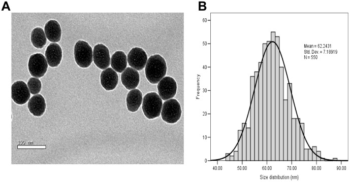
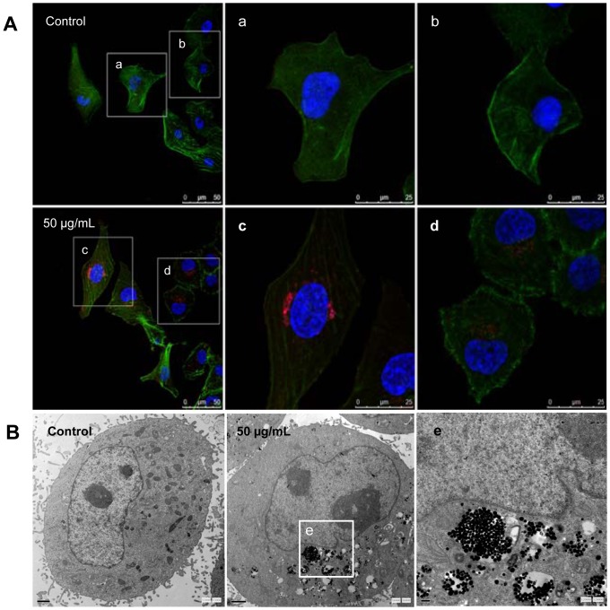
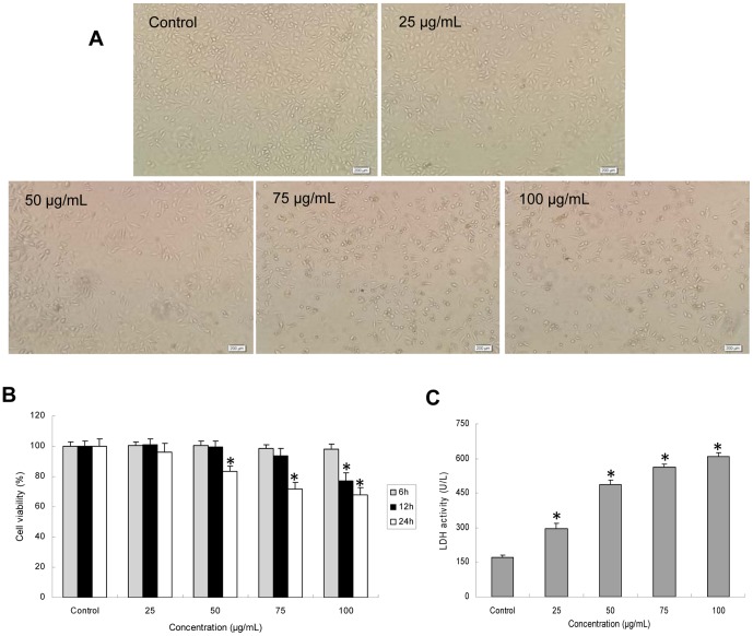
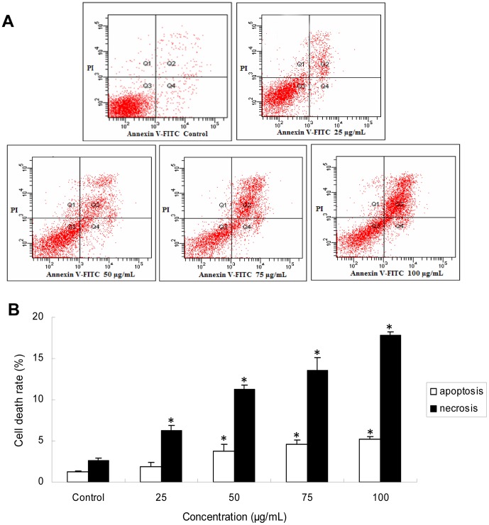
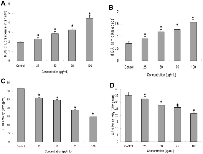
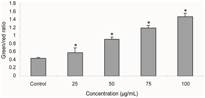
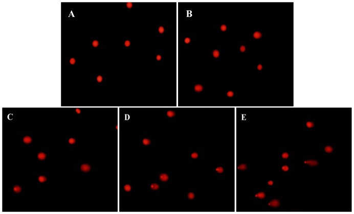
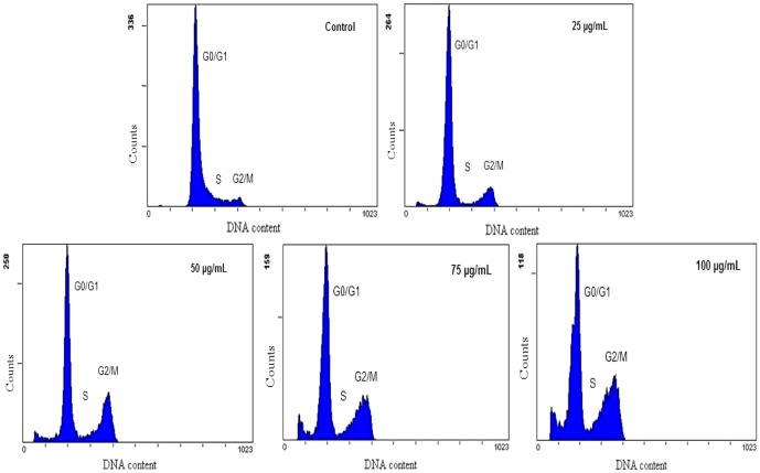
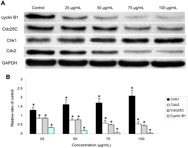
Silica nanoparticles have become promising carriers for drug delivery or gene therapy. Endothelial cells could be directly exposed to silica nanoparticles by intravenous administration. However, the underlying toxic effect mechanisms of silica nanoparticles on endothelial cells are still poorly understood. In order to clarify the cytotoxicity of endothelial cells induced by silica nanoparticles and its mechanisms, cellular morphology, cell viability and lactate dehydrogenase (LDH) release were observed in human umbilical vein endothelial cells (HUVECs) as assessing cytotoxicity, resulted in a dose- and time- dependent manner. Silica nanoparticles-induced reactive oxygen species (ROS) generation caused oxidative damage followed by the production of malondialdehyde (MDA) as well as the inhibition of superoxide dismutase (SOD) and glutathione peroxidase (GSH-Px). Both necrosis and apoptosis were increased significantly after 24 h exposure. The mitochondrial membrane potential (MMP) decreased obviously in a dose-dependent manner. The degree of DNA damage including the percentage of tail DNA, tail length and Olive tail moment (OTM) were markedly aggravated. Silica nanoparticles also induced G2/M arrest through the upregulation of Chk1 and the downregulation of Cdc25C, cyclin B1/Cdc2. In summary, our data indicated that the toxic effect mechanisms of silica nanoparticles on endothelial cells was through DNA damage response (DDR) via Chk1-dependent G2/M checkpoint signaling pathway, suggesting that exposure to silica nanoparticles could be a potential hazards for the development of cardiovascular diseases.
Conflict of interest statement
Figures
Similar articles
-
RETRACTED: Combined Effect of Silica Nanoparticles and Benzo[a]pyrene on Cell Cycle Arrest Induction and Apoptosis in Human Umbilical Vein Endothelial Cells.Int J Environ Res Public Health. 2017 Mar 9;14(3):289. doi: 10.3390/ijerph14030289. Int J Environ Res Public Health. 2017. Retraction in: Int J Environ Res Public Health. 2025 Mar 27;22(4):508. doi: 10.3390/ijerph22040508. PMID: 28282959 Free PMC article. Retracted.
-
Cardiovascular toxicity evaluation of silica nanoparticles in endothelial cells and zebrafish model.Biomaterials. 2013 Jul;34(23):5853-62. doi: 10.1016/j.biomaterials.2013.04.032. Epub 2013 May 8. Biomaterials. 2013. PMID: 23663927
-
Oxidative stress-mediated mitochondrial pathway-dependent apoptosis is induced by silica nanoparticles in H9c2 cardiomyocytes.Toxicol Mech Methods. 2020 Nov;30(9):646-655. doi: 10.1080/15376516.2020.1805664. Epub 2020 Aug 20. Toxicol Mech Methods. 2020. PMID: 32746757
-
Molybdenum nanoparticles-induced cytotoxicity, oxidative stress, G2/M arrest, and DNA damage in mouse skin fibroblast cells (L929).Colloids Surf B Biointerfaces. 2015 Jan 1;125:73-81. doi: 10.1016/j.colsurfb.2014.11.014. Epub 2014 Nov 20. Colloids Surf B Biointerfaces. 2015. PMID: 25437066
-
Genotoxicity evaluation of silica nanoparticles in murine: a systematic review and meta-analysis.Toxicol Mech Methods. 2022 Jan;32(1):1-17. doi: 10.1080/15376516.2021.1965277. Epub 2021 Aug 25. Toxicol Mech Methods. 2022. PMID: 34350812
Cited by
-
Global Proteomics to Study Silica Nanoparticle-Induced Cytotoxicity and Its Mechanisms in HepG2 Cells.Biomolecules. 2021 Mar 2;11(3):375. doi: 10.3390/biom11030375. Biomolecules. 2021. PMID: 33801561 Free PMC article.
-
Cellular Alterations Due to Direct and Indirect Interaction of Nanomaterials with Nucleic Acids.Int J Mol Sci. 2024 Feb 6;25(4):1983. doi: 10.3390/ijms25041983. Int J Mol Sci. 2024. PMID: 38396662 Free PMC article. Review.
-
Silica nanoparticles induce reversible damage of spermatogenic cells via RIPK1 signal pathways in C57 mice.Int J Nanomedicine. 2016 May 24;11:2251-64. doi: 10.2147/IJN.S102268. eCollection 2016. Int J Nanomedicine. 2016. PMID: 27307728 Free PMC article.
-
Evaluation of the DNA damaging potential of indigenous health hazardous quartz nanoparticles on the cultured lung cells.Toxicol Res (Camb). 2016 Dec 20;6(2):152-161. doi: 10.1039/c6tx00414h. eCollection 2017 Mar 1. Toxicol Res (Camb). 2016. PMID: 30090485 Free PMC article.
-
Threshold Dose of Three Types of Quantum Dots (QDs) Induces Oxidative Stress Triggers DNA Damage and Apoptosis in Mouse Fibroblast L929 Cells.Int J Environ Res Public Health. 2015 Oct 26;12(10):13435-54. doi: 10.3390/ijerph121013435. Int J Environ Res Public Health. 2015. PMID: 26516873 Free PMC article.
References
-
- Li Z, Barnes JC, Bosoy A, Stoddart JF, Zink JI (2012) Mesoporous silica nanoparticles in biomedical applications. Chem Soc Rev 41: 2590–605. - PubMed
-
- Lee JE, Lee N, Kim T, Kim J, Hyeon T (2011) Multifunctional mesoporous silica nanocomposite nanoparticles for theranostic applications. Acc Chem Res 44: 893–902. - PubMed
-
- Kang JH, Keller JJ, Chen CS, Lin HC (2012) Asian dust storm events are associated with an acute increase in pneumonia hospitalization. Ann Epidemiol 22: 257–263. - PubMed
Publication types
MeSH terms
Substances
LinkOut - more resources
Full Text Sources
Other Literature Sources
Research Materials
Miscellaneous
