. 2013 Apr 19;8(4):e62199.
doi: 10.1371/journal.pone.0062199. Print 2013.

Role of sensory experience in functional development of Drosophila motor circuits

Affiliations

Role of sensory experience in functional development of Drosophila motor circuits

Akira Fushiki et al. PLoS One. .

Abstract

Neuronal circuits are formed according to a genetically predetermined program and then reconstructed in an experience-dependent manner. While the existence of experience-dependent plasticity has been demonstrated for the visual and other sensory systems, it remains unknown whether this is also the case for motor systems. Here we examined the effects of eliminating sensory inputs on the development of peristaltic movements in Drosophila embryos and larvae. The peristalsis is initially slow and uncoordinated, but gradually develops into a mature pattern during late embryonic stages. We tested whether inhibiting the transmission of specific sensory neurons during this period would have lasting effects on the properties of the sensorimotor circuits. We applied Shibire-mediated inhibition for six hours during embryonic development (15-21 h after egg laying [AEL]) and studied its effects on peristalsis in the mature second- and third-instar larvae. We found that inhibition of chordotonal organs, but not multidendritic neurons, led to a lasting decrease in the speed of larval locomotion. To narrow down the sensitive period, we applied shorter inhibition at various embryonic and larval stages and found that two-hour inhibition during 16-20 h AEL, but not at earlier or later stages, was sufficient to cause the effect. These results suggest that neural activity mediated by specific sensory neurons is involved in the maturation of sensorimotor circuits in Drosophila and that there is a critical period for this plastic change. Consistent with a role of chordotonal neurons in sensory feedback, these neurons were activated during larval peristalsis and acute inhibition of their activity decreased the speed of larval locomotion.


Conflict of interest statement

Competing Interests: The authors have declared that no competing interests exist.

Figures

Figure 1
Figure 1. Behavioral analysis (locomotion analysis).
To measure larval propagation duration, we videotaped the animals’ behavior using a CCD camera (30 frames/sec for 30 seconds). We manually calculated the duration of waves (10 waves per larva) using ImageJ software.
Figure 2
Figure 2. Inhibition of chos at late embryonic stage affects peristalsis in mature larvae.
Effects of inhibiting the activity of multidendritic neurons (md neurons, mds) or chordotonal organs (chos) during 15–21 h AEL. Inhibition of chos (orange boxes) but not md neurons (blue boxes) led to propagation defects in the second (A) and third (B) instar. Box plots indicate the median value (horizontal line inside the box), 25–75% quartiles (box), and the data range (whiskers). Statistical significance was determined by Mann-Whitney U test (**P<0.01; n.s., not significant). For all conditions, n = 10. RT, restrictive temperature. 109(2)80-GAL4 and iav-GAL4 were used as md neurons and chos drivers, respectively.
Figure 3
Figure 3. Chordotonal organs regulate locomotion through sensory feedback to the CNS.
(A, B) Increase in propagation duration in iav1 mutants (A) and in larvae expressing TeTxLc (TTX) in chos (B). (C) Acute inhibition of chos with Shibirets increased the propagation duration of the larvae. Statistical analysis was done by Welch’s t test (***P<0.001; *P<0.05; n.s., not significant). For all conditions, n = 10. RT, restrictive temperature. Controls were as follows: yw (A), yw>UAS-TTX (B), yw>UAS-Shi (C).
Figure 4
Figure 4. Calcium imaging in chordotonal organs during muscle contractions.
GCaMP-based calcium imaging in chos. (A) Representative fluorescence change (ΔF/F0) of GCaMP3 in the axon terminals of chos in a posterior (ROI1, blue) and an anterior (ROI2, red) region of the CNS were plotted (bottom). Note that the signal rise in the posterior region precedes that in the anterior region in consecutive rounds of activity propagation. The amplitude of calcium signals was smoothed (moving average of 60 points). The top panel shows the position of the ROIs. (B) Time-lapse images of the fluorescent intensities (t1 to t4). Anterior is to the left and posterior is to the right. Arrows denote the signal rise in chos terminals in the ventral nerve cord. Arrowheads denote positions of muscle contractions, which were detected using the autofluorescence images of muscles. Note that the activation of chos and segmental muscle contraction propagate at a similar timing (see Movie S1). Scale bars represent 250 µm.
Figure 5
Figure 5. A critical period for the maturation of the sensorimotor circuit.
(A, C) The effects of temporal inhibition of chos neurons at various embryonic stages on larval locomotion, analyzed at the second (A, 48 h AEL) and third instar (C, 96 h AEL) stage. A two-hour temperature shift to a restricted temperature was applied to iav-GAL4>UAS-Shits embryos at various stages during embryogenesis. The temperature shift at specific embryonic periods increased the propagation duration. (B, D) The same temperature shift applied to control embryos (yw>UAS-Shits ) had no effect on locomotion, when analyzed at the second (B) and third instar (D) stage. Statistical significance was determined by one-way ANOVA followed by Dunnett’s test for multiple comparisons (**P<0.01; *P<0.05; n.s., not significant compared with iav-GAL4>UAS-Shits (none) control). For all conditions, n = 10. RT, restrictive temperature.
Figure 6
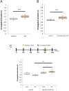
Figure 6. Inhibition of chordotonal organs at larval stage has no effect on the speed of larval locomotion.
In contrast to the two-hour inhibition during the embryonic stage (17–19 h AEL), those during first (A, 32–34 h AEL) and third (B, 70–72 h AEL) instar stage had no effect on the speed of locomotion. Analyzed at second (A, 48 h AEL) and third instar (B, 96 h AEL) stages. Statistical significance was determined by one-way ANOVA followed by Tukey’s test for multiple comparisons (***P<0.001; **P<0.01). For all conditions, n = 10. RT, restrictive temperature.

Similar articles

Cited by

References

    1. Brown TG (1911) The intrinsic factors in the act of progression in the mammal. Proceedings of the Royal Society of London Series B-Containing Papers of a Biological Character 84: 308–319.
    1. Grillner S, Wallen P (1985) Central pattern generators for locomotion, with special reference to vertebrates. Annu Rev Neurosci 8: 233–261. - PubMed
    1. Buschges A (2005) Sensory control and organization of neural networks mediating coordination of multisegmental organs for locomotion. J Neurophysiol 93: 1127–1135. - PubMed
    1. Buonomano DV, Merzenich MM (1998) Cortical plasticity: from synapses to maps. Annu Rev Neurosci 21: 149–186. - PubMed
    1. Hubel DH, Wiesel TN (1970) The period of susceptibility to the physiological effects of unilateral eye closure in kittens. J Physiol 206: 419–436. - PMC - PubMed

Publication types

LinkOut - more resources