. 2013 Apr 19;8(4):e62251.
doi: 10.1371/journal.pone.0062251. Print 2013.

TnaA, an SP-RING protein, interacts with Osa, a subunit of the chromatin remodeling complex BRAHMA and with the SUMOylation pathway in Drosophila melanogaster

Affiliations

TnaA, an SP-RING protein, interacts with Osa, a subunit of the chromatin remodeling complex BRAHMA and with the SUMOylation pathway in Drosophila melanogaster

Juan Monribot-Villanueva et al. PLoS One. .

Abstract

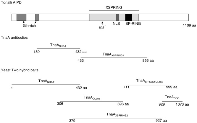
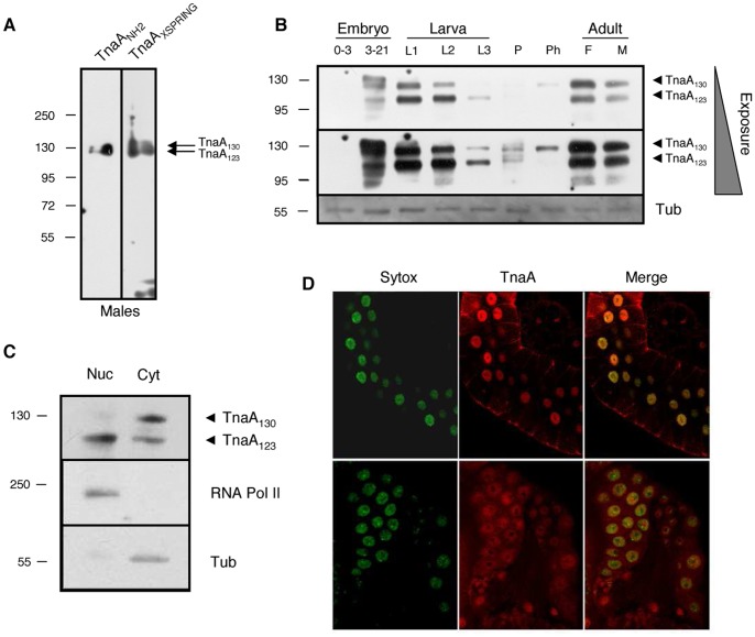
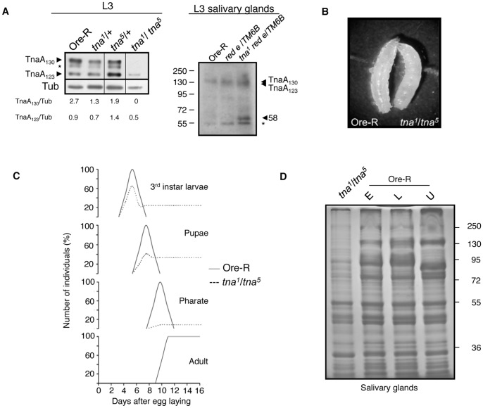
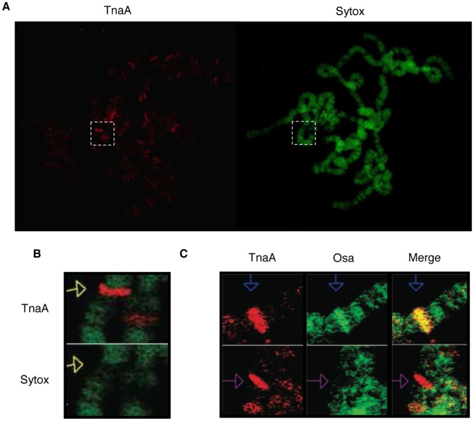
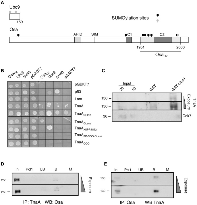
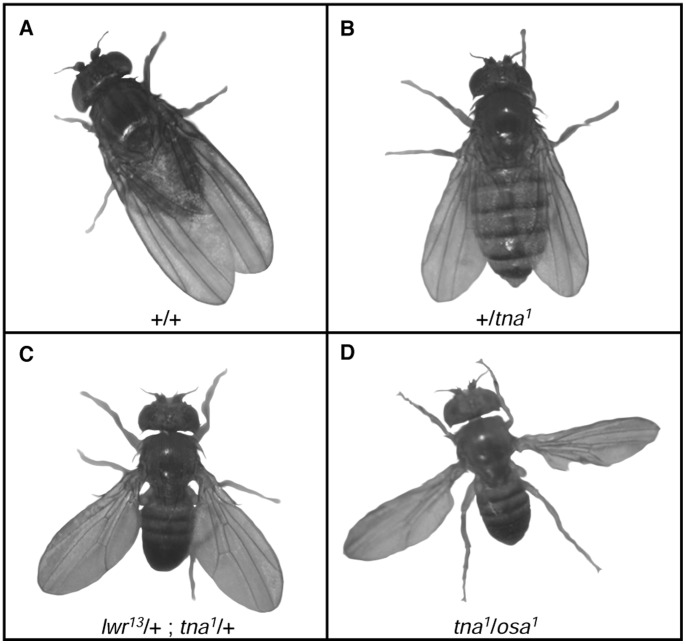
Tonalli A (TnaA) is a Drosophila melanogaster protein with an XSPRING domain. The XSPRING domain harbors an SP-RING zinc-finger, which is characteristic of proteins with SUMO E3 ligase activity. TnaA is required for homeotic gene expression and is presumably involved in the SUMOylation pathway. Here we analyzed some aspects of the TnaA location in embryo and larval stages and its genetic and biochemical interaction with SUMOylation pathway proteins. We describe that there are at least two TnaA proteins (TnaA130 and TnaA123) differentially expressed throughout development. We show that TnaA is chromatin-associated at discrete sites on polytene salivary gland chromosomes of third instar larvae and that tna mutant individuals do not survive to adulthood, with most dying as third instar larvae or pupae. The tna mutants that ultimately die as third instar larvae have an extended life span of at least 4 to 15 days as other SUMOylation pathway mutants. We show that TnaA physically interacts with the SUMO E2 conjugating enzyme Ubc9, and with the BRM complex subunit Osa. Furthermore, we show that tna and osa interact genetically with SUMOylation pathway components and individuals carrying mutations for these genes show a phenotype that can be the consequence of misexpression of developmental-related genes.


Conflict of interest statement

Competing Interests: The authors have declared that no competing interests exist.

Figures

Figure 1
Figure 1. TnaA protein, domains and fragments.
The TnaA protein (upper section). TnaA domains are indicated. Nuclear localization signal is NLS. The stop codon in the tna1 allele is indicated by an arrow. TnaA fragments used to produce TnaA antibodies from GST fusion proteins are shown (middle section). TnaA fragments fused to the yeast GAL4-binding domain to use as baits in two-hybrid assays (lower section).
Figure 2
Figure 2. Expression and location of TnaA proteins throughout Drosophila development.
(A) TnaANH2 and TnaAXSPRING antibodies detect the same proteins. TnaA proteins detected by full-range Western analysis in an adult male soluble protein extract with TnaANH2 and TnaAXSPRING antibodies (1∶100 dilution). (B) TnaA developmental Western. Detection of TnaA130 and Tna123 isoforms in soluble extracts isolated from embryos (0–3 and 3–21 hour), 1st, 2nd and 3rd instar larvae (L1, L2 and L3), pupae (P), pharate (Ph) and female and male adults (F and M). β-tubulin was used as a protein loading control (bottom). The TnaAXSPRING and β-tubulin antibodies were used 1∶100 and 1∶1000, respectively. (C) TnaA123 is mainly nuclear. Detection of TnaA proteins in nuclear (Nuc) and cytoplasmic (Cyt) soluble fractions isolated from embryos 3–21 hour. The largest RNA polymerase II subunit and β-tubulin were used as controls of nuclear and cytoplasmic fractions, respectively. TnaANH2, RNA polymerase II, and β-tubulin antibodies were used 1∶120, 1∶500, and 1∶1000, respectively. (D) Immunostaining of TnaA in salivary (upper panel) and ring glands (lower panel) of Ore-R third instar larvae with TnaAXSPRING (3∶5, red), DNA (Sytox, green) and merge (yellow). We detected no signal when immunostaining was done with secondary antibody only (not shown).
Figure 3
Figure 3. Characterization of tna1/tna5 individuals.
(A) TnaA proteins in tna1/tna5 third instar larvae (left panel). Note the absence of TnaA130 and the reduction of TnaA123 levels. Western blot probed with the TnaAXSPRING antibody (1∶100) was done with similar amount of proteins from total third instar larvae extracts of the indicated genotype. The band marked with an asterisk (*) appears sometimes depending on extracts and running gel conditions and may represent a processed protein product. Narrow vertical lines indicate edition of lanes from the same film with samples from flies with other tna genotypes not relevant to this work. β-tubulin was used as loading control (antibody used 1∶1000). The levels of TnaA130 and TnaA123 were compared with the level of β-tubulin using Image-J. Western blot of tna1 red e/TM6B salivary gland extracts probed with TnaANH2 antibody (right panel) detected TnaA130, TnaA123 (that comigrated in this gel) and a 58 kDa band close to the predicted size of a truncated Tna-1 protein (62 kDa). Salivary gland extracts from OreR individuals and from individuals harbouring the tna1 parental (red e) and balancer (TM6B) chromosomes were used as controls. The asterisk indicates a crossreacting band present in all the genotypes tested. (B) tna1/tna5 and Ore-R third instar larvae. (C) Survival percentage of tna1/tna5 individuals at different stages of development. Ore-R (−) and tna1/tna5 (–) survival percentages are indicated. More than 100 third instar larvae were counted for each genotype. Heterozygous tna individuals have survival rates similar to those for Ore-R. (D) Protein profile of tna1/tna5 salivary glands. Note the differences between the tna1/tna5 and the Ore-R total protein profiles. SDS-PAGE of soluble protein extracts from 10 pairs of larval salivary glands stained with Coomasie blue. Ore-R salivary glands extracts from early (E) and late (L) or unstaged (U) third instar larvae that were loaded as references.
Figure 4
Figure 4. TnaA is located on polytene salivary gland chromosomes of third instar larvae and sometimes colocalizes with Osa.
(A) Immunostaining of TnaA in Ore-R (wild type) polytene salivary gland chromosomes of third instar larvae. TnaAXSPRING antibody (1∶50, red) and DNA (Sytox, green). Amplification in B is indicated (pointed white rectangle). (B) TnaA is located in chromatin interbands. (C) TnaA and Osa colocalize in some sites on polytene salivary gland chromosomes of third instar larvae (blue arrows in the top panels) but in others do not (purple arrows in the bottom panels). TnaAXSPRING antibody (1∶50, red) and Osa (1∶50, green). No signal was detected when no primary antibody was added (data not shown).
Figure 5
Figure 5. TnaA interacts with Drosophila Ubc9 and with Osa.
(A) Schemes of Drosophila Ubc9 and OsaC2 used in biochemical assays. In the Osa protein, the ARID, the C1 and C2 domains (grey boxes), the SUMO interacting motif (SIM) and the OsaC2 fragment (dark line) are indicated. Forward (black circles) and inverted (gray circles) putative SUMOylation consensus sites in these proteins are indicated. For TnaA baits see Fig. 1. (B) TnaA interaction with Ubc9 and OsaC2 in yeast two-hybrid assays. Yeast colony complementation of growth controls in SD-Trp/−Leu media due to the presence of pGBKT7 (Trp+) and pGADT7, (Leu+) plasmids (left) in the same yeast cells. Interaction assay in QDO +3-AT (SD-Trp/−Leu/−Ade/−His +3-AT) media (right). Growth is observed when baits and preys interact, allowing GAL4 reconstitution with the consequent ADE2 and HIS3 reporter genes transcription. Baits were TnaA fragments (Fig. 1) fused to the DNA-binding domain of GAL4 in pGBKT7. Ubc9 and OsaC2 were preys fused to the GAL4 activation domain in pGADT7. Human p53 (p53) and Lamin C (Lam) interactions with SV40 are positive and negative controls, respectively. (C) TnaA interaction with Ubc9 by pull-down. The assays were done with 10 µg of each GST or GST-Ubc9 as baits and with 500 µg of soluble nuclear fraction from 3–21 hour embryos. 10 and 20% of the extract are shown as Input. TnaA was detected by Western analysis with TnaAXSPRING antibody (1∶100) when GST-Ubc9 was used as bait. The 130 kDa weight marker is indicated (left) and increasing exposures of the same membrane are shown. Cdk7 was detected only in the Input lanes (antibody dilution, 1∶1000). (D) Coimmunoprecipitation of Osa with TnaA antibodies from nuclear extracts obtained from 3–21 hour embryos. TnaAXSPRING antibodies (1 µg), and 3–21 hour embryos soluble nuclear fraction (500 µg) were used. The Western was revealed with the Osa antibody (1∶1000). Input, (In), preclearing 1 (Pcl1), unbound (Ub), bound (B). Immunoprecipitation with the equivalent amount of a preimmune serum instead of TnaAXSPRING antibody was used as Mock (M). Both panels show films with increasing exposure time of the same membrane. (E) Coimmunoprecipitation of TnaA with Osa antibodies from total extracts obtained from 3–21 hour embryos. Osa antibodies (1 µg), and 3–21 hour embryos soluble nuclear fraction (3.7 mg) were used. The Western was revealed with the TnaANH2 antibody (1∶120). Lanes are labeled as above. The equivalent amount of an irrelevant antibody was used as mock (M). Molecular weight markers are indicated (left).
Figure 6
Figure 6. SUMOylation pathway mutations enhance held-out wing phenotype of tna and osa flies.
Flies with different held-out wing phenotype expressivity. Fly genotype is indicated in each picture. Penetrance of the held-out wing phenotype in each genotype is in Table 1 (A) Wild type fly (B) Slight held-out wing phenotype of +/tna1 flies. The same phenotype is presented by +/osa1 individuals. (C) Stronger held-out wing phenotype of smt3/+;tna1/+ individuals. The same phenotype is presented by lwr/+;tna1/+ or smt304493/+;osa1/+ individuals. (D) Strongest held-out wing phenotype of tna1/osa1 individuals. This phenotype is also presented by tna1/osa2 individuals.

Similar articles

Cited by

References

    1. Geiss-Friedlander R, Melchior F (2007) Concepts in SUMOylation: a decade on. Nat Rev Mol Cell Biol 10: 947–956. - PubMed
    1. Rodríguez MS, Dargemont C, Hay RT (2001) SUMO-1 conjugation in vivo requires both a consensus modification motif and nuclear targeting. J Biol Chem 276: 12654–12659. - PubMed
    1. Nie M, Xie Y, Loo JA, Courey AJ (2009) Genetic and proteomic evidence for roles of Drosophila SUMO in cell cycle control, Ras signaling, and early pattern formation. PLoS One 4: e5905. - PMC - PubMed
    1. Gareau JR, Lima CD (2010) The SUMO pathway: emerging mechanisms that shape specificity, conjugation and recognition. Nat Rev Mol Cell Biol 11: 861–871. - PMC - PubMed
    1. Gutiérrez L, Zurita M, Kennison JA, Vázquez M (2003) The Drosophila trithorax group gene tonalli (tna) interacts genetically with the Brahma remodeling complex and encodes an SP-RING finger protein. Development 130: 343–354. - PubMed

Publication types

MeSH terms

LinkOut - more resources